

Method for extracting exosome from chicken bile and application of exosome in immunology
A technology of extracellular body and chicken bile, applied in the field of immunology, can solve the problem of no report on the extracellular body of avian source bile, and achieve the effects of high uniformity, good shape retention and short time consumption.
- Summary
- Abstract
- Description
- Claims
- Application Information
AI Technical Summary
Problems solved by technology
Method used
Image

Examples
Embodiment 1
[0048] Embodiment 1, the separation and purification of extracellular body in chicken bile
[0049] 1. Extraction of extracellular bodies in chicken bile: Extract bile from SPF chicken gallbladder within 4 hours of isolation, mix it with PBS buffer in equal volume, centrifuge at 2000g for 15min, 4000g for 30min, and 8000g for 45min at a low temperature of 4°C. Centrifuge at 15,000 g for 60 min to remove cell debris, take the supernatant and filter it through a filter membrane (through double-layer filter paper, 5 μm filter membrane, 1.2 μm filter membrane, and 0.22 μm filter membrane in turn), centrifuge at 110,000 g at 4°C for 2 h, and resuspend the precipitate with PBS. Then centrifuge at 110,000 g at 4°C for 90 min and dissolve the precipitate with PBS, which is the extracted extracellular body.
[0050] In this method, the precipitate after centrifugation at 110,000g at 4°C for 90 minutes can be further separated by density gradient centrifugation with 30wt%, 45wt%, and 60...
Embodiment 2
[0053] Embodiment 2, identification of extracellular body in chicken bile
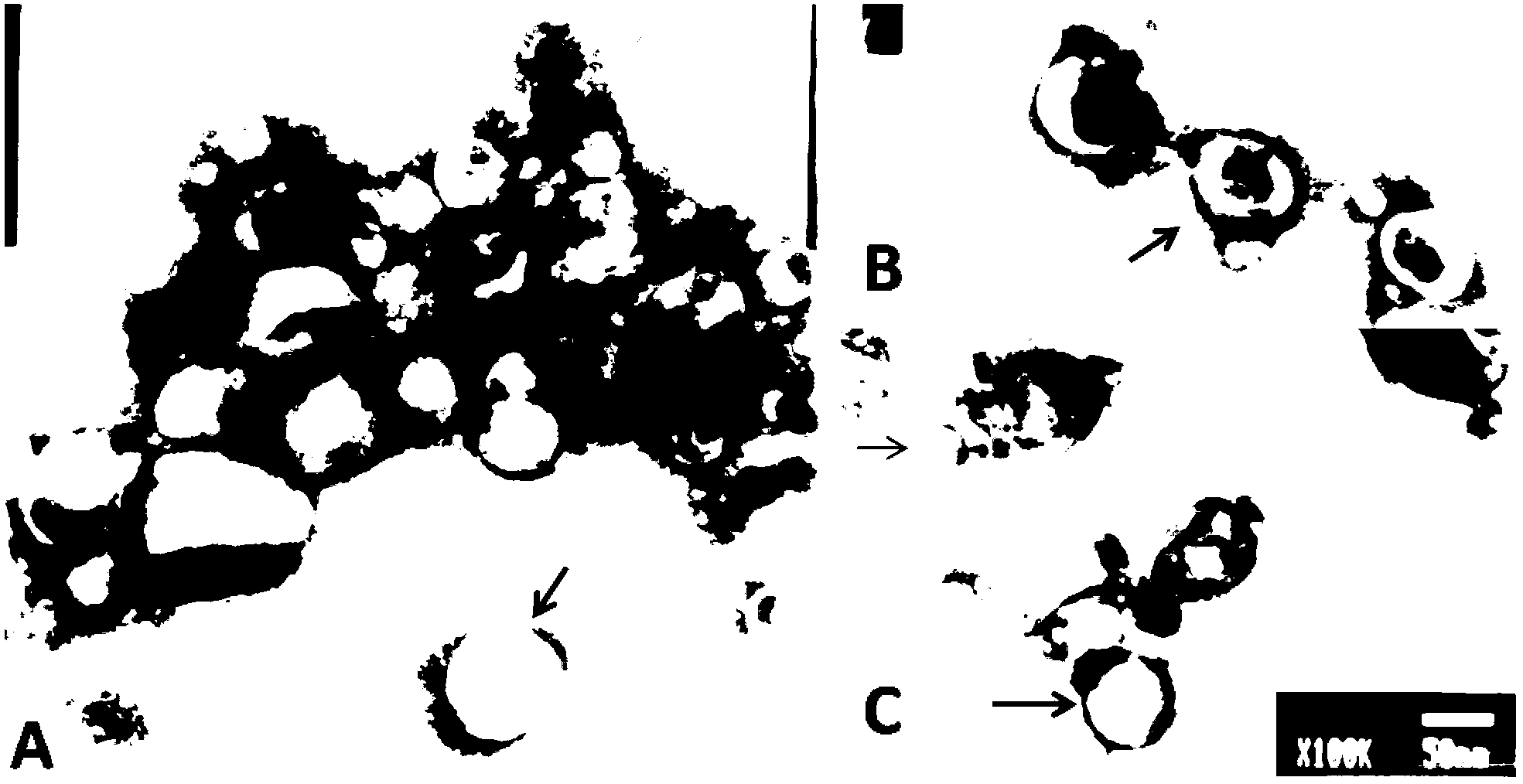
[0054] 1. Morphological observation of exosomes
[0055] Take 10 μL of the extracellular body stock solution on the sample-loading copper grid, and let it stand at room temperature for 1 min; blot the liquid from the side with filter paper, add about 10 μL of 1% uranyl acetate dropwise on the copper grid, and negatively stain at room temperature for 1 min; blot the negative with filter paper The dye solution was washed once with four distilled water, dried under an incandescent lamp, observed and photographed under a transmission electron microscope.
[0056] Such as figure 1 As shown in A-C, the exosomes in chicken bile are small vesicles with a typical cup-shaped structure with a diameter of 30-150 nm.
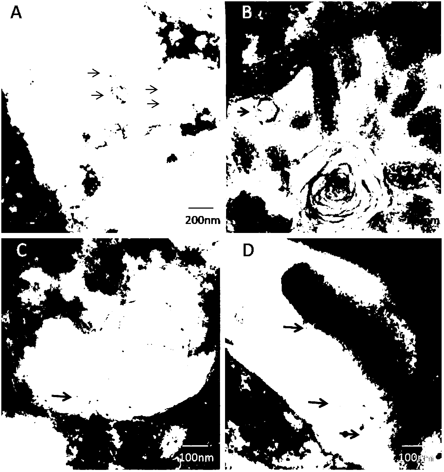
[0057] 2. Observation of Exosome Secretion and Release
[0058] (1) Sample processing: take the liver tissue of SPF chicken within 1 min of isolation, and trim it into strips with a size of 1mm×1m...
Embodiment 3
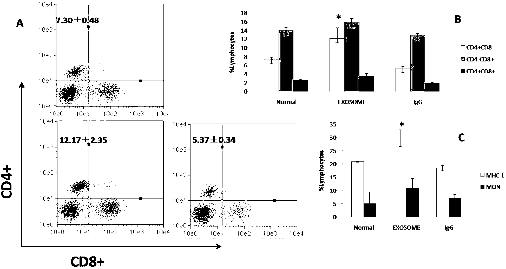
[0075] Example 3, Exosome MHC Correlation Detection
[0076] 1. Pre-processing:
[0077] Take the extracellular body suspension and place it in 100 μl of 4mm acetaldehyde / sulfate magnetic bead emulsion (purchased from Invitrogen), incubate at 20°C for 15min, then add PBS buffer and shake gently on the shaker for 2h, and finally add 100mmol / L glycine Stop the reaction.
[0078] 2. Flow Cytometry Analysis
[0079] (1) Centrifuge the exosomes connected to the magnetic beads at 2000rpm for 5min,
[0080] (2) Disperse the cells by blowing with 1ml Facs Buffer, centrifuge at 2000rpm for 5min, pour out the buffer,
[0081] (3) Add 100 microliters of Facs Buffer to each tube
[0082] (4) Take a certain amount of samples from each tube to form three tubes for comparison
[0083] (5) Centrifuge at 2000rpm for 5min,
[0084] (6) Use chicken antibody CD4 + T-FITC / CD8 + T-PE, MHC Ⅰ-FITC / MON-PE and other antibodies were stained at 4°C in the dark for 30 minutes.
[0085] (7) Wash w...
PUM
| Property | Measurement | Unit |
|---|---|---|
| Diameter | aaaaa | aaaaa |
| Mass density | aaaaa | aaaaa |
Abstract
Description
Claims
Application Information