Multi-epitope fusion antigen for detecting virus serum antibody of porcine reproductive and respiratory syndrome and kit prepared with multi-epitope fusion antigen

A technology for respiratory syndrome and fusion antigen, applied in biological testing, hybrid peptides, material inspection products, etc., can solve the problems of time-consuming, complicated and laborious virus purification process, and achieve long response duration, simple purification process, and control The effect of missed detection risk

Active Publication Date: 2012-10-17
重庆业为基生物科技集团有限公司
View PDF5 Cites 4 Cited by
  • Summary
  • Abstract
  • Description
  • Claims
  • Application Information

AI Technical Summary

Problems solved by technology

[0006] The gene structure and antigenicity of porcine reproductive and respiratory syndrome virus are relatively complex, and have the characteristics of antigenic variability, which makes it difficult to achieve ideal sensitivity for single antigen detection or antibody detection through a single antigen
The advantage of using the whole virus as a coated antigen is that the antigen production method is relatively simple. The disadvantage is that the virus purification process is complicated, time-consuming, laborious, and costly. The ELISA detection established by the whole virus antigen has strong background and non-specific reactions. There is a large difference in batches, and there is a hidden danger of poisoning, etc.
Although the use of complete virus particles as a standard antigen can have ideal sensitivity, and the antigen production method is relatively simple, it requires complex processes such as virus culture, inactivation, and verification after inactivation, which is time-consuming, laborious, and costly. Whole virus The ELISA established by antigen has strong background reaction and non-specific reaction, and the difference between different batches is large, which has the hidden danger of potential virus transmission risk, etc. These shortcomings make this method not suitable for industrial development; in addition, the complete Virus particles are used as antigens to detect antibodies, which are also prone to false positive results

Method used


Image

  • Multi-epitope fusion antigen for detecting virus serum antibody of porcine reproductive and respiratory syndrome and kit prepared with multi-epitope fusion antigen
  • Multi-epitope fusion antigen for detecting virus serum antibody of porcine reproductive and respiratory syndrome and kit prepared with multi-epitope fusion antigen
  • Multi-epitope fusion antigen for detecting virus serum antibody of porcine reproductive and respiratory syndrome and kit prepared with multi-epitope fusion antigen

Examples


Embodiment 1

[0051] Example 1 Design of multiple epitope fusion antigen (MEFA) NSP7-N1 and cloning of its coding gene

[0052] 1. Design of multiple epitope fusion antigen NSP7-N1

[0053] According to the online database search provided by NCBI, the genome sequence of porcine reproductive and respiratory syndrome virus (GenBank: JQ663567.1), the amino acid residue sequence of NSP7 (NCBI Reference Sequence: NP 740601.1), and the amino acid residue sequence of nucleocapsid protein N can be obtained (GenBank: AAY53878.1). Wherein NSP7 is encoded by ORF1a nucleotide residues, divided according to the non-structural proteins of European porcine reproductive and respiratory syndrome virus Lelystad strain, the start and end positions of the Nsp7 amino acid coding sequence are Ser2083-Glu2351. The starting and ending positions of the Nsp7 amino acid sequence of North American porcine reproductive and respiratory syndrome virus VR-2332 strain are Ser2200-Glu2458. The porcine reproductive and res...

Embodiment 2

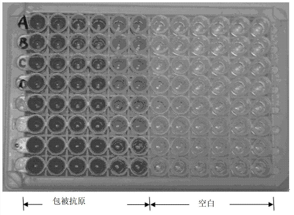
[0057] Transform BL21codon plus (DE3) engineering bacteria by conventional methods (molecular cloning: an experimental manual (New York: Cold Spring Harbor Laboratory Press, 1989)), take the recombinant pET42a-NSP7-N1 plasmid to transform BL21codon plus (DE3) bacteria, and spread on the Chloramphenicol-resistant and kanamycin-resistant LB solid medium, cultivated overnight in a 37°C incubator, picked white colonies and inoculated them in LB medium for expanded culture the next day, cultured at 30°C, and the measured bacterial OD value reached 0.6 At ~0.8 hours, IPTG with a final concentration of 0.6mmol / L was added to induce, and the induced bacteria were collected at time points 1, 2, 4, and 6 hours for SDS-PAGE identification. The results showed that the engineered bacteria induced by IPTG had a specific protein band with a molecular weight of about 34kD, which was consistent with the expected molecular weight value of the expression product of pET42a-NSP7-N1, accounting for ...

Embodiment 3

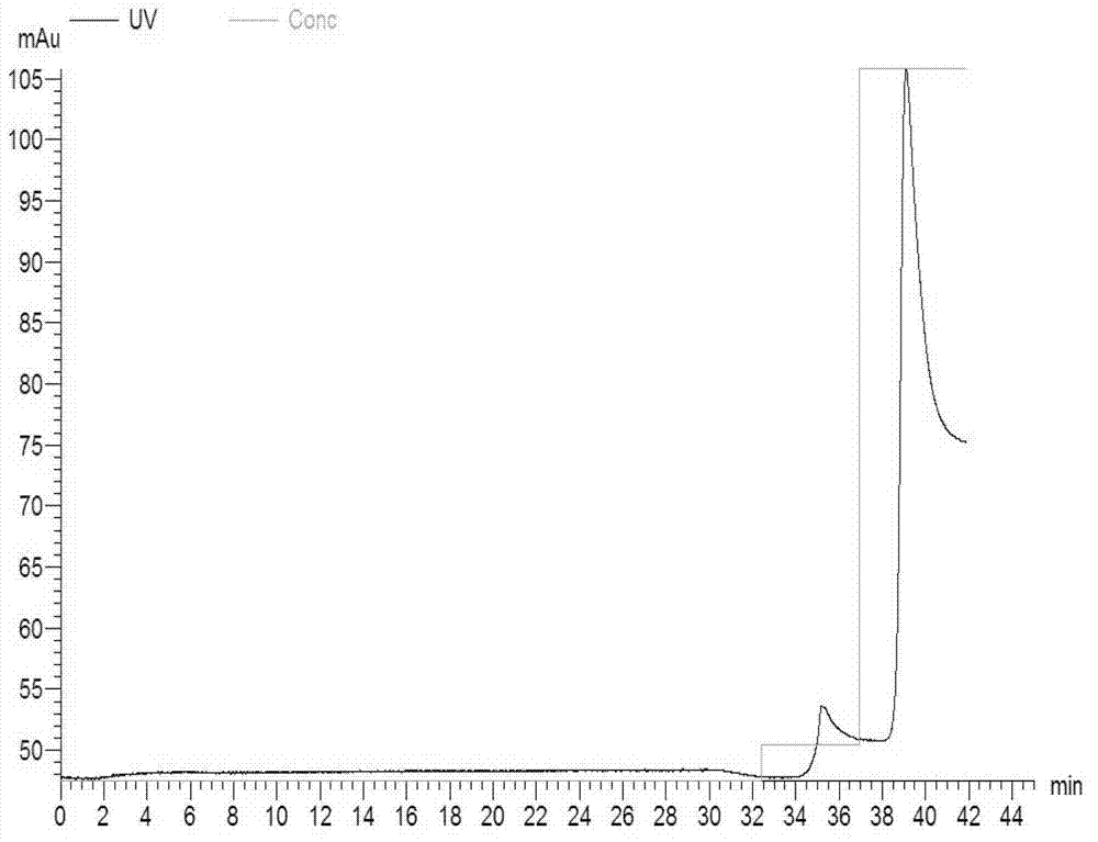
[0058] The purification of embodiment 3NSP7-N1 recombinant protein

[0059] The engineered bacteria transformed with the pET42a-NSP7-N1 recombinant plasmid were induced and expressed by 0.6mmol / L IPTG, and then purified using GSTrap F.F. After suspension, the bacteria were ultrasonically broken on ice, centrifuged at 4°C at high speed, and the supernatant was filtered and put on the column with Amersham's AKTAprime. , pH8.0) to collect the eluted protein. Dilute the eluted peak containing the target protein with His-binding buffer (20mmol / l, sodium phosphate, 0.5M NaCl, pH7.4) at a volume ratio of 1:9 and then repurify on HisTrap HP, His-binding buffer After equilibration, use His-elution buffer (20mmol / L sodium phosphate, 0.5M NaCl, 0.5M imidazole, pH7.4), first elute with 10% buffer to remove non-specific binding impurities, and then use 100% buffer after equilibration solution to wash off the target protein. A desalting column is used to desalt and remove imidazole, and ...


PUM

PropertyMeasurementUnit
diameteraaaaaaaaaa

Abstract

The invention relates to a multi-epitope fusion antigen for detecting virus serum antibody of porcine reproductive and respiratory syndrome, wherein an amino acid sequence of the multi-epitope fusion antigen is shown in SEQ ID No:1. The invention also relates to a preparation method of the multi-epitope fusion antigen, and a kit for detecting the virus antibody of porcine reproductive and respiratory syndrome. The multi-epitope fusion antigen prepared by the preparation method has the advantages of high purity, good stability, simple preparation process, and low production cost. The kit has the characteristics of high sensitivity and strong safety.

Description

technical field [0001] The invention relates to a multi-epitope fusion antigen for detecting porcine reproductive and respiratory syndrome virus serum antibody and a kit for its preparation. Background technique [0002] Porcine reproductive and respiratory syndrome (PRRS), commonly known as porcine blue ear disease, is a new porcine infectious disease that emerged in the late 1980s, and its pathogen is porcine reproductive and respiratory syndrome virus (Porcine reproductive and respiratory syndrome virus) and respiratory syndrome virus, PRRSV). The occurrence of the disease was first reported in the United States in 1987. Its symptoms are mainly manifested as severe reproductive disorders such as abortion, premature birth, stillbirth and mummification in pregnant sows, increased respiratory symptoms and mortality before weaning in piglets, dyspnea in bred pigs, developmental Sluggishness, loss of libido in boars, decline in semen quality, etc. A devastating epidemic brok...

Claims


Application Information

Patent Timeline
Patent Type & Authority Applications(China)
IPC IPC(8): C07K19/00C12N15/62C12N15/63C12N1/21C12N1/19C12N5/10G01N33/68
Inventor 黄洪涛严俊姚静石延宾张宪胡伟江雨丽李小鱼魏勇
Owner 重庆业为基生物科技集团有限公司
Why Eureka
Try Eureka