Novel bionic surface-enhanced Raman spectrum base and preparation method thereof

A surface-enhanced Raman and substrate technology, applied in the field of Raman spectroscopy and nanomaterials, can solve the problems of low hot spot density of the substrate, difficulty in large-scale production, complicated manufacturing process, etc., and achieve convenient material acquisition, strong stability, and simple processing Effect

Inactive Publication Date: 2013-12-18
HUAZHONG AGRI UNIV
View PDF1 Cites 17 Cited by
  • Summary
  • Abstract
  • Description
  • Claims
  • Application Information

AI Technical Summary

Problems solved by technology

However, the equipment required by this method is expensive, the preparation cost is high, and the prepared substrate has low hot spot density and general enhancement effect.
Emerging 3D (three-dimensional) substrates, such as silicon nanopillar arrays modified with noble metal nano

Method used


Image

  • Novel bionic surface-enhanced Raman spectrum base and preparation method thereof
  • Novel bionic surface-enhanced Raman spectrum base and preparation method thereof
  • Novel bionic surface-enhanced Raman spectrum base and preparation method thereof

Examples


Embodiment 1

[0025] Example 1: Preparation of gold-coated two-dimensional biomimetic surface-enhanced Raman spectroscopy substrate

[0026] 1) Raw material pretreatment: select clean and transparent black grasshopper (Cryptotympana atrata Fabricius) forewings, ultrasonically clean them with ultrapure water for 5 minutes, dry them in the air, remove the wing veins with a scalpel and cut them into 4×4 squares Then use tweezers to fix it on a glass slide with double-sided adhesive tape, blow it flat with an ear wash ball, and use it as a biological template.

[0027] 2) Substrate preparation: Put the prepared biological template into a DC ion sputtering apparatus (ETD-3000), use high-purity argon (99.999%) as the protective gas, and use high-purity gold (99.99%) as the target material, and sputter The air pressure is 10 Pa, the sputtering current is 4 mA, the sputtering rate is 3.0-4.0 nanometers per minute, preferably 3.6-4.0 nanometers per minute, using intermittent sputtering (one minute o...

Embodiment 2

[0029] Example 2: Preparation of silver-coated two-dimensional biomimetic surface-enhanced Raman spectroscopy substrate

[0030] 1) Pretreatment of raw materials: same as step 1) in Example 1.

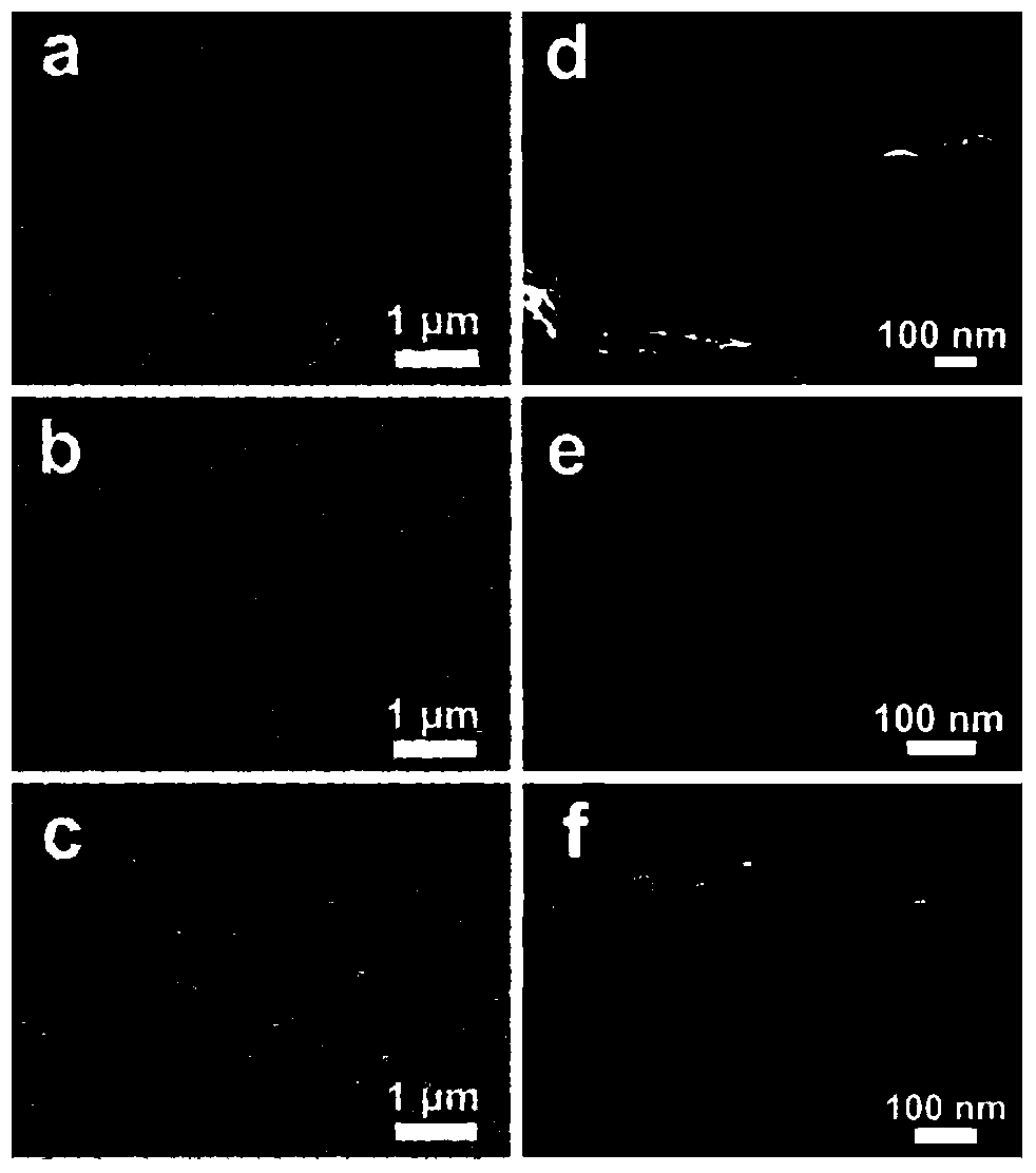
[0031] 2) Substrate preparation: the sputtering conditions and methods are the same as those described in step 2) in Example 1. The gold layer is intermittently sputtered for 2 cycles, and then the high-purity silver (99.99%) target is used, and the microstructure of the silver-coated layer can be adjusted by sputtering for 12 to 14 cycles. Such as figure 2 As shown in b and 2e, after 13 cycles of silver sputtering, a columnar two-dimensional biomimetic surface-enhanced Raman spectroscopy substrate with a silver-coated gap of less than 10 nm (ie, a two-dimensional silver-coated substrate) was prepared. In contrast, under the conditions of sputtering pressure of 6 Pa and sputtering current of 6 mA, 8 cycles of intermittent sputtering can also prepare pure silver-coated two-dimensiona...

Embodiment 3

[0033] Example 3: Preparation of silver particle-modified three-dimensional biomimetic surface-enhanced Raman spectroscopy substrate

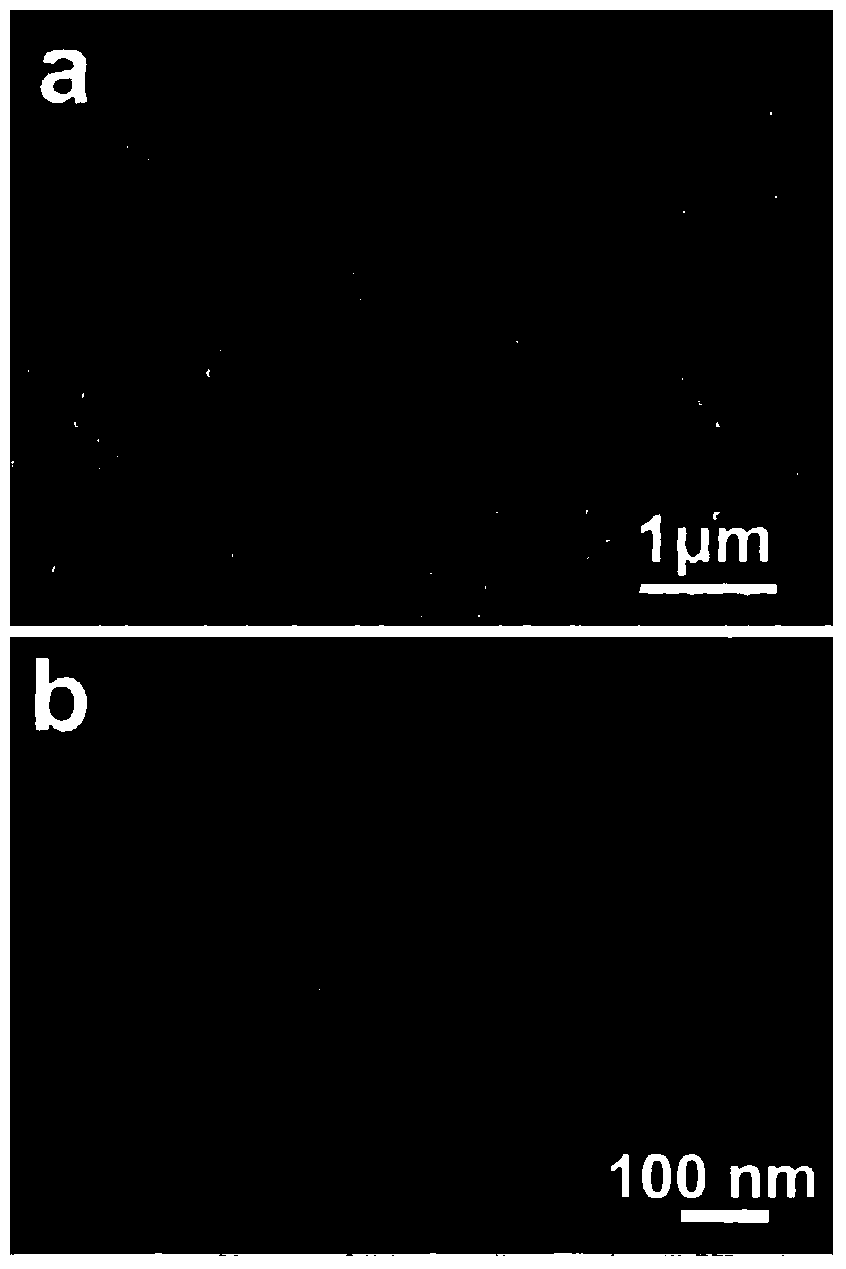
[0034] 1) Pretreatment of raw materials: same as step 1) in Example 1. Because the surface of the cicada wing of the black grasshopper has quasi-regular chitin nano-columnar array knots, its chitin columns are wide at the top and narrow at the bottom, with a diameter of about 60 nanometers at the top, a diameter of about 120 nanometers at the bottom, and a height of about 180 nanometers. The gap between them is about 80 nm. This special microstructure facilitates the formation of silver particles during the preparation of silver particle-modified 3D biomimetic surface-enhanced Raman spectroscopy substrates.

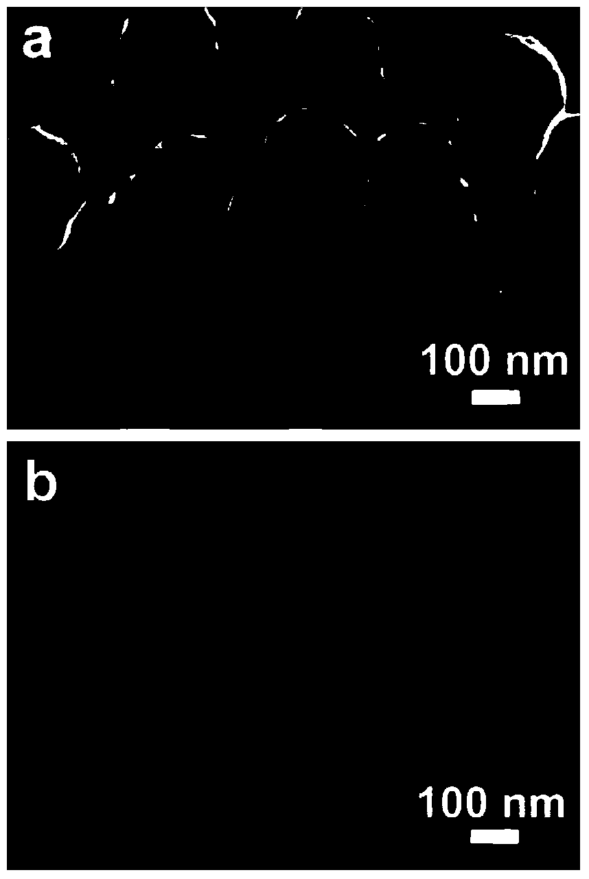
[0035] 2) Substrate preparation: the sputtering conditions and methods are the same as those described in step 2) in Example 1. The microstructure of the silver nanoparticle layer can be adjusted by direct sputtering with the silver targ...


PUM

PropertyMeasurementUnit
Thicknessaaaaaaaaaa
Bottom diameteraaaaaaaaaa

Abstract

The invention discloses a novel bionic surface-enhanced Raman spectrum base and a preparation method thereof. The base is prepared by the following steps: 1) pre-treating raw materials, namely ultrasonically cleaning the cicada wings, removing the veins, cutting into small blocks, fixing and flattening to obtain a biological template; 2) preparing the base, namely placing the biological template obtained in the step 1) in a direct current ion sputtering instrument, sputtering and depositing precious metal on the biological template in an intermittent sputtering mode by using high-pure precious metal as a target under an inert atmosphere, so as to obtain the novel bionic surface-enhanced Raman spectrum base, wherein the high-pure precious metal is gold and/or silver with the purity not less than 99.99%; the intermittent sputtering mode means that stopping lasts for 45-75s after sputtering lasting for 45-75s, the sputtering process and the stopping process are combined as a period. The two-dimensional or three-dimensional prepared by using the cicada wings which are abundant in source, convenient for material taking and simple in treatment as the template is clean in surface, high in enhancement factor, good in reproducibility and strong in stability.

Description

technical field [0001] The invention belongs to the technical fields of Raman spectroscopy and nanometer materials, and in particular relates to a novel bionic surface-enhanced Raman spectroscopy substrate and a preparation method thereof. Background technique [0002] Since its discovery in the mid-1970s, surface-enhanced Raman scattering (SERS) technology has been widely used in biomedicine due to its unique advantages such as narrow spectral band, high sensitivity, anti-photobleaching, in-situ, non-destructive, and providing fingerprint information. , surface science, molecular recognition, trace detection and many other fields have been more and more widely used. The surface-enhanced Raman spectroscopy substrate can significantly increase the Raman signal of the molecule through the electromagnetic field enhancement effect, so that the Raman signal of the molecule adsorbed on the enhanced substrate is about 10 times stronger than that of the ordinary molecule. 4 -10 7 ...

Claims


Application Information

Patent Timeline
IPC IPC(8): C23C14/34C23C14/20G01N21/65
Inventor 韩鹤友邵锋吕志成
Owner HUAZHONG AGRI UNIV
Why Eureka
Try Eureka