
Production method of model for retinitis pigmentosa disease of primate

A primate, retinal pigment technology, applied in the field of making a primate retinitis pigmentosa disease model, can solve the problems of damage to animal rights, waste of experimental animals, increase the difficulty of experiments, etc., to achieve high modeling success rate, guarantee Effects of animal welfare, avoidance of systemic injury

Active Publication Date: 2014-04-16
JIANGSU PROVINCE HOSPITAL
View PDF1 Cites 2 Cited by
  • Summary
  • Abstract
  • Description
  • Claims
  • Application Information

AI Technical Summary

Problems solved by technology

Primates are most similar to humans in structure and function, and primate disease models are extremely important for the study of human diseases, but in primates, intravenous injection of a corresponding dose of sodium iodate cannot achieve satisfactory retinitis pigmentosa In the model, increasing the dose increases the systemic toxicity of the drug, and the animals even die due to the systemic toxicity of the drug
The application number is 201010131148.0, and the Chinese patent titled "Rhesus monkey retinal degeneration model and its drug screening method" discloses a method for preparing a rhesus monkey retinal degeneration model by intravenous injection of sodium iodate, but using intravenous injection of sodium iodate For modeling, the drug first flows back to the heart through the vein, and then is pumped out through the heart through the aorta, carotid artery, and ophthalmic artery to enter the retina-choroidal circulation. The effect of the pigment epithelium is concentration-dependent. In order to achieve the purpose of destroying the pigment epithelium, the dosage of the drug should be increased, but this will correspondingly increase the systemic toxicity of the drug
Our pre-experiment shows that the lethal dose of the drug is close to the model-making dose by intravenous injection of sodium iodate. The ERG function recovered spontaneously in half a month, and systemic side effects, hematuria, acute kidney injury, etc. were common, and the modeling rate was low, and the mortality rate was high, which increased the difficulty of the experiment, wasted experimental animals, and damaged animal rights, so it could not be used satisfactorily. experiment

Method used


Image

  • Production method of model for retinitis pigmentosa disease of primate
  • Production method of model for retinitis pigmentosa disease of primate
  • Production method of model for retinitis pigmentosa disease of primate

Examples


Embodiment 1

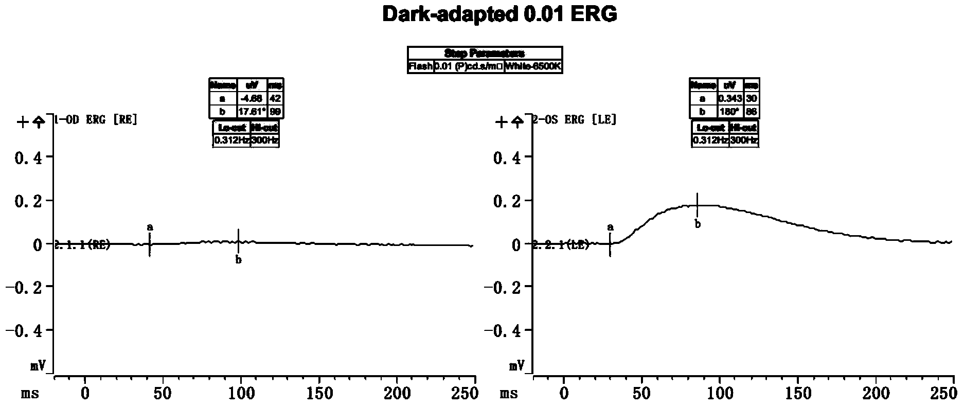
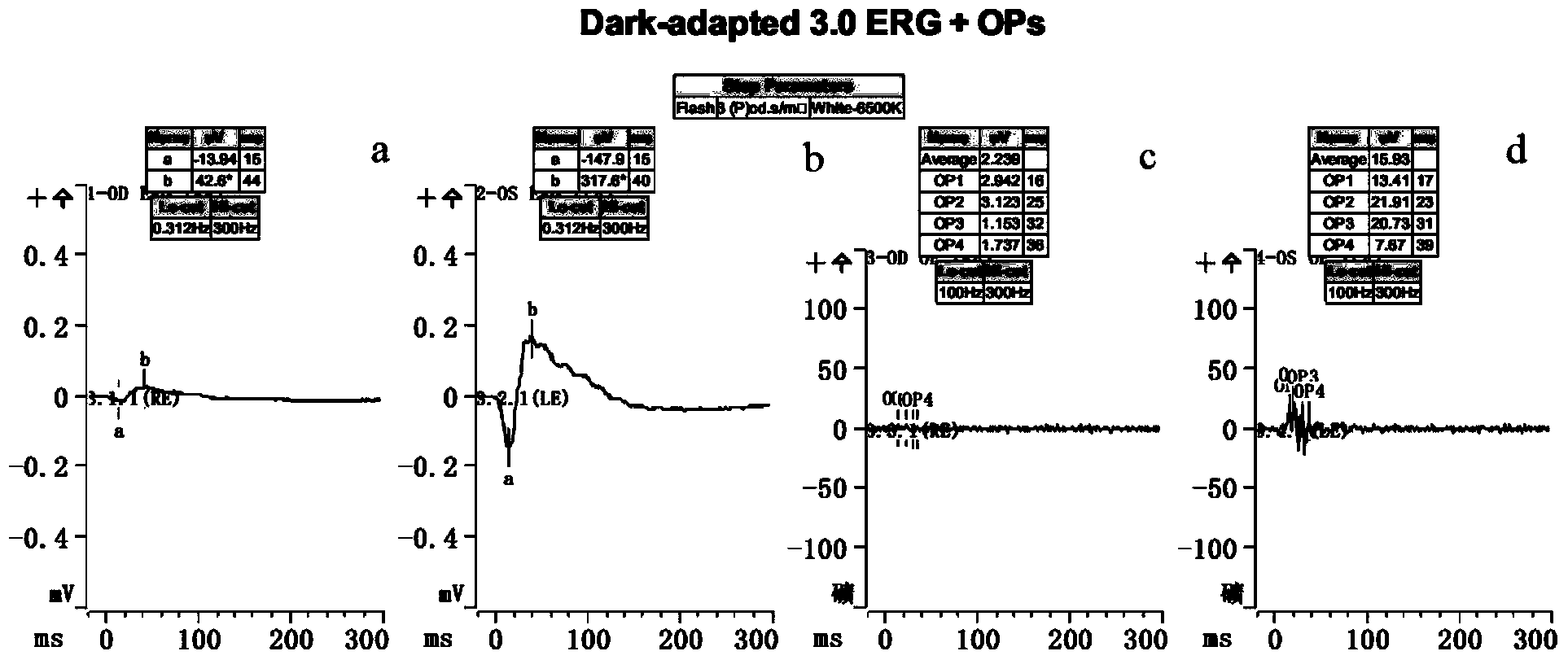
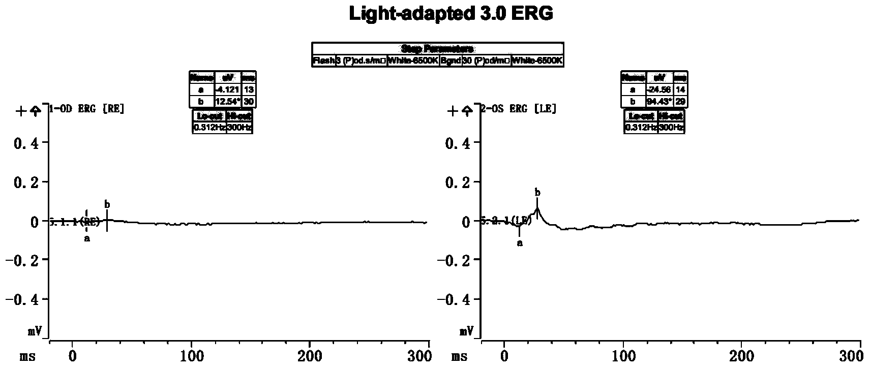
[0030] Example 1 A model of retinitis pigmentosa in cynomolgus monkeys was established by injecting sodium iodate into the carotid artery.

[0031] Objective: To establish a model of retinitis pigmentosa in cynomolgus monkeys by injecting sodium iodate into the carotid artery, and to screen out the appropriate dose for modeling.

[0032] Experimental Materials:

[0033] Main reagents:

[0034] Anesthetics: 50mg / ml ketamine hydrochloride, 3% pentobarbital sodium

[0035] Disinfection medicine: 5.0g / L povidone-iodine

[0036] Modeling drugs: sodium iodate (Sigma, batch number: S4007), in a dark room environment, the sodium iodate powder was melted to a concentration of 10 mg / ml with 0.9% sterile saline, and filtered with a 0.22 μm filter.

[0037] Fundus fluorescence contrast agent: sodium fluorescein injection (Lishede) 500mg / 5ml / bottle

[0038] Main instruments:

[0039] YZ5F slit lamp microscope (Hangzhou Liuliu)

[0040] Espion Visual Electrophysiology Apparatus (Diagn...


PUM


Abstract

The invention discloses a production method of a model for retinitis pigmentosa disease on a primate. A certain dosage of sodium iodate is applied to the primate by a way of carotid artery injection. The production method disclosed by the invention is easy and feasible, high in success rate of model production, good in repeatability, and stable in model; the needed drug amount of sodium iodate is less, so that the damage of a large dosage of sodium iodate to the whole body of the animal is avoided, and the animal lives for a long time so as to provide possibility for follow-up tests; the model for any one side or two sides can be produced according to needs; the production of the model for one side cannot cause any damage to a fellow eye so as to ensure the eyesight of the animal for normal life; the self-control study on bilateral eyes can be carried out on one animal so as to provide the credibility of the tests, and reduce the number of required experimental animals. The production method disclosed by the invention provides a good tool for the research on the retinitis pigmentosa, and has important significance for both pathophysiology research and treatment research of the disease.

Description

technical field [0001] The invention relates to the field of medical technology, in particular to a method for making a primate animal retinitis pigmentosa disease model. Background technique [0002] The retinal pigment epithelium is very important to maintain normal retinal functions. Retinitis Pigmentosa (RP), which belongs to cone and rod cell dystrophy, is a group of hereditary diseases. Calmness and photoreceptor dysfunction are characterized. The possible pathogenesis is due to the phagocytosis of the retinal pigment epithelial cells to the outer segmental disc of the visual cell, and the decline in digestion, resulting in the residue of the disc disintegration and the formation of a layer of obstacles, which hinders the rotation of nutrients from the choroid to the retina, thereby causing Progressive dystrophy and gradual degeneration and apoptosis of visual cells. Today, there is still no clear and effective treatment, and its pathogenesis and treatment methods ha...

Claims


Application Information

Patent Timeline
IPC IPC(8): A61K33/18
Inventor 计江东袁松涛刘庆淮
Owner JIANGSU PROVINCE HOSPITAL
Learn More