Method and oligonucleotide for detecting FGFR3 gene G380R site mutation

An oligonucleotide and site mutation technology, which is applied in the field of highly sensitive detection of gene FGFR3G380R site mutation, can solve the problem of long distance between reporter fluorophore and quencher fluorophore, failure to recognize double-stranded DNA sequence, fluorescence quenching Incomplete extinction and other problems, to achieve the effect of long detection cycle, short detection cycle, simplified design and cost

Active Publication Date: 2014-05-14
杭州艾迪康医学检验中心有限公司
View PDF1 Cites 2 Cited by
  • Summary
  • Abstract
  • Description
  • Claims
  • Application Information

AI Technical Summary

Problems solved by technology

Among them, SYBR Green I is not as specific as the double-hybrid probe method and Taqman probe method because it is an unsaturated dye, and its specificity must be judged by observing the melting curve. In addition, it cannot recognize a specific double-stranded DNA sequence, as long as it is double-stranded DNA The sequence will be combined with luminescence, so the background is usually high, and false positives may occur when used clinically
The two-hybridization probe method involves two specific probes that are complementary to the template and adjacent (distance 1-5bp), the 3' end of the upstream probe is labeled with a donor fluorophore, and the 5' end of the adjacent downstream probe is End-labeled Red640 acceptor fluorophore, the cost is relatively expensive
The molecular beacon method involves a stem-loop double-labeled oligonucleotide probe in a hairpin structure. The nucleic acid sequences at both ends of the probe are complementary paired, and a fluorescent group labeled at one end is labeled with a quencher group at the other end. The biggest disadvantage of this method is that the probe cannot be completely combined with the template during hybridization, so the stability is poor, and the labeling process is more complicated when the probe is synthesized.
The Taqman probe method involves a gene-specific probe (usually 20-30bp long) complementary to the template. The 5' end and the 3' end of the probe are respectively labeled with a reporter fluorophore and a quencher fluorophore, but The longer probe sequence in this method makes the distance between the reporter fluorophore and the quencher fluorophore at both ends of the probe farther away, resulting in incomplete fluorescence quenching, and the quencher group will also produce fluorescence of different wavelengths. This will make the background higher

Method used


Image

  • Method and oligonucleotide for detecting FGFR3 gene G380R site mutation
  • Method and oligonucleotide for detecting FGFR3 gene G380R site mutation
  • Method and oligonucleotide for detecting FGFR3 gene G380R site mutation

Examples


Embodiment 1

[0045] Example 1 Oligonucleotides and kits for detecting mutations at the G380R site of the FGFR3 gene

[0046] The oligonucleotide used to detect the mutation of the G380R site of the FGFR3 gene in the sample, the oligonucleotide includes a pair of specific amplification products SEQ NO1 and SEQ NO2, and its base sequence is:

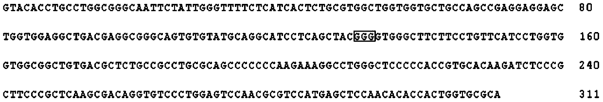
[0047] SEQ NO 1: GGGTTTTTCTCATCACTCTGCG

[0048] SEQ NO2: AGTGTGTATGCAGGCATCCT,

[0049] And a pair of specific detection probes SEQ NO3 and SEQ NO4, its base sequence is:

[0050] SEQ NO 3: FAM-AAGAAGCCCACCCCGTAGCT-MGB

[0051] SEQ NO4: HEX-AAGAAGCCCACCCTGTAGCT-MGB

[0052] Among them, MGB is marked at the 3' end of SEQ NO3 and SEQ NO4 to enhance detection specificity.

[0053] A kit for detecting mutations at the G380R site of the FGFR3 gene in a sample, the kit including sample DNA extraction reagents, erythrocyte lysate, absolute ethanol, qPCR amplification reaction solution, positive control substance, negative control substance and blank cont...

Embodiment 2

[0063] Embodiment 2: detection process

[0064] (1) Blood DNA extraction: Take 500ul whole blood, put it into a 1.5ml centrifuge tube, add 1ml red blood cell lysate. Turn it upside down to make it completely mixed, rotate and pulsate for 15 seconds, and then put it into a centrifuge for centrifugation at 5000rpm for 10min. Pour off the upper layer, and it can be seen that there is a bloody precipitate at the bottom of the centrifuge tube. Add 500ul red blood cell lysate, repeat this lysis step once. Centrifuge at 5000rpm for 5min, and finally suck up all the upper layer with a pipette and discard it, so that the blood-colored precipitate at the bottom of the centrifuge tube no longer has lysate. Mix well with a metal bath at 100°C for 10 minutes, 12000 rpm for 5 minutes, take the supernatant, and obtain the sample DNA solution. When not used immediately, the sample DNA solution was stored at -20°C until use.

[0065] (2) Real-time fluorescent PCR amplification:

[0066] T...

Embodiment 3

[0070] Embodiment 3: Sample detection verification

[0071] 7 clinical samples were selected, numbered 1-7, among which samples 1-5 were clinically diagnosed as patients with ACH, sample 6 was from the wife of patient 5, the wife of patient 5 did not suffer from ACH, and had been pregnant for 20 weeks, and sample 7 was number 5 Amniotic fluid from the patient's wife. The implementation steps are as follows:

[0072] (1) Samples 1-6 are peripheral blood. DNA was extracted according to the blood DNA extraction method described in Example 2. Sample 7 was extracted from chorion exfoliated cells in amniotic fluid using the QIAamp DNA extraction kit from QIAGEN.

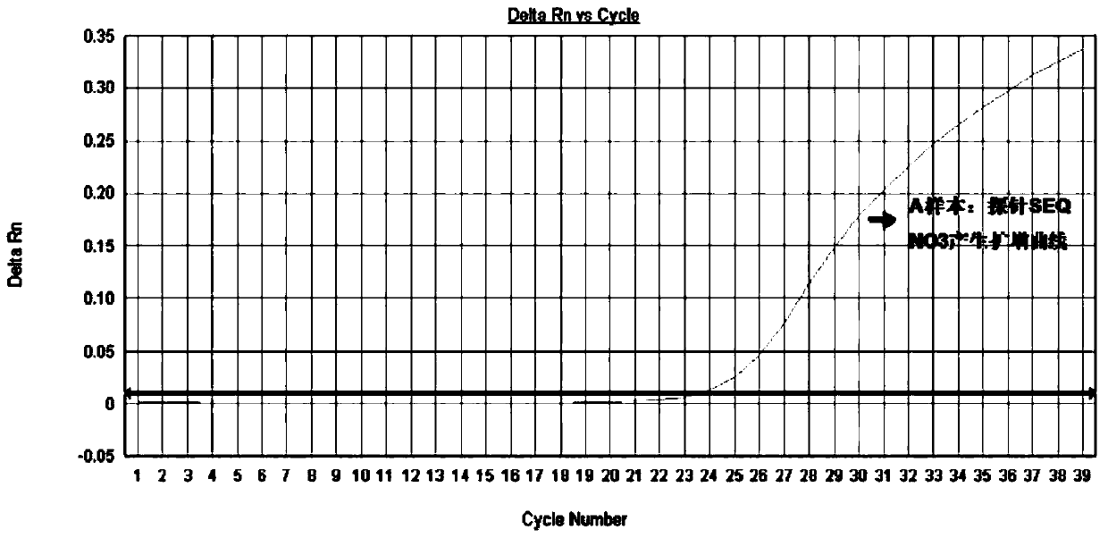
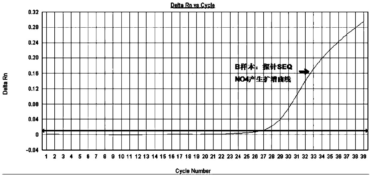
[0073] (2) According to the real-time fluorescent PCR amplification method described in Example 2, real-time fluorescent PCR detection was performed on the DNA of samples 1-7, and Sanger sequencing analysis was performed on the 10th exon of the FGFR3 gene of these 7 samples. The results of real-time fluorescent PCR (qPCR...


PUM


Abstract

The invention discloses a method and oligonucleotide for detecting FGFR3 gene G380R site mutation, and relates to a pair of specific amplification primers SEQ NO 1 and SEQ NO 2, and a pair of specific detection probes SEQ NO 3 and SEQ NO 4. By adopting the method and oligonucleotide, congenital achondroplasia can be rapidly diagnosed and identified, and the method and oligonucleotide have the advantages of being short in detection cycle, good in specificity, high in accuracy, high in sensitivity, less in condition dependence, low in pollution risk and the like.

Description

technical field [0001] The invention belongs to the field of gene mutation detection, in particular to a method and oligonucleotide for highly sensitive detection of gene FGFR3G380R site mutation. Background technique [0002] Fibroblast growth factor receptor 3 (FGFR3), belonging to the fibroblast growth factor receptors (FGFRs) family, is a transmembrane protein that regulates development and other functions, and plays an important role in bone development . The FGFR3 gene is located on human chromosome 4p1613 and contains 806 amino acid residues. It is a tyrosine kinase receptor and consists of an extracellular ligand-binding region modified by glycosylation, a hydrophobic transmembrane region and a Composition of the catalytic domain of intracellular tyrosine kinases. The FGFR3 gene is about 1615kb in length, with 19 exons and 18 introns, of which the 10th exon encodes the transmembrane region of FGFR3. [0003] Achondroplasia (ACH) is a common hereditary disease of t...

Claims


Application Information

Patent Timeline
Patent Type & Authority Applications(China)
IPC IPC(8): C12Q1/68C12N15/11
CPCC12Q1/686C12Q2527/107
Inventor 徐建成孙翠莲
Owner 杭州艾迪康医学检验中心有限公司
Why Eureka
Try Eureka