Fusion protein containing leucine-rich repetitive sequence, and preparation method and application thereof

A technology of fusion protein and protein, applied in the field of fusion protein, can solve the problems of many impurities, inaccurate expression of target protein, cumbersome process and steps, etc.

Inactive Publication Date: 2014-10-29
李华顺
View PDF2 Cites 4 Cited by
  • Summary
  • Abstract
  • Description
  • Claims
  • Application Information

AI Technical Summary

Problems solved by technology

Usually, proteins with histidine can be adsorbed by nickel agarose gel or nickel NTA agarose gel under natural conditions, but because the His tag is very small, if it is folded into the protein during the fusion protein folding process, the His tag will be caused If it is not easy to expose, it is difficult to carry out protein purification; at the same time, the His tag does have the problem of

Method used


Image

  • Fusion protein containing leucine-rich repetitive sequence, and preparation method and application thereof
  • Fusion protein containing leucine-rich repetitive sequence, and preparation method and application thereof
  • Fusion protein containing leucine-rich repetitive sequence, and preparation method and application thereof

Examples


Embodiment 1

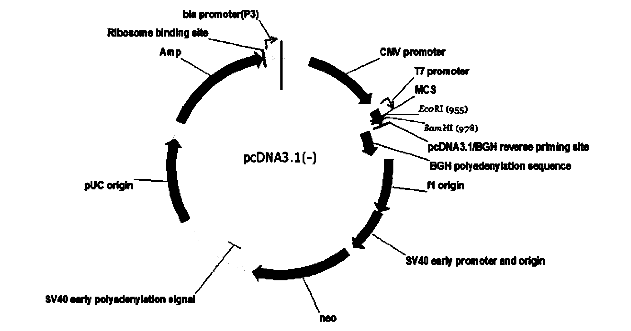
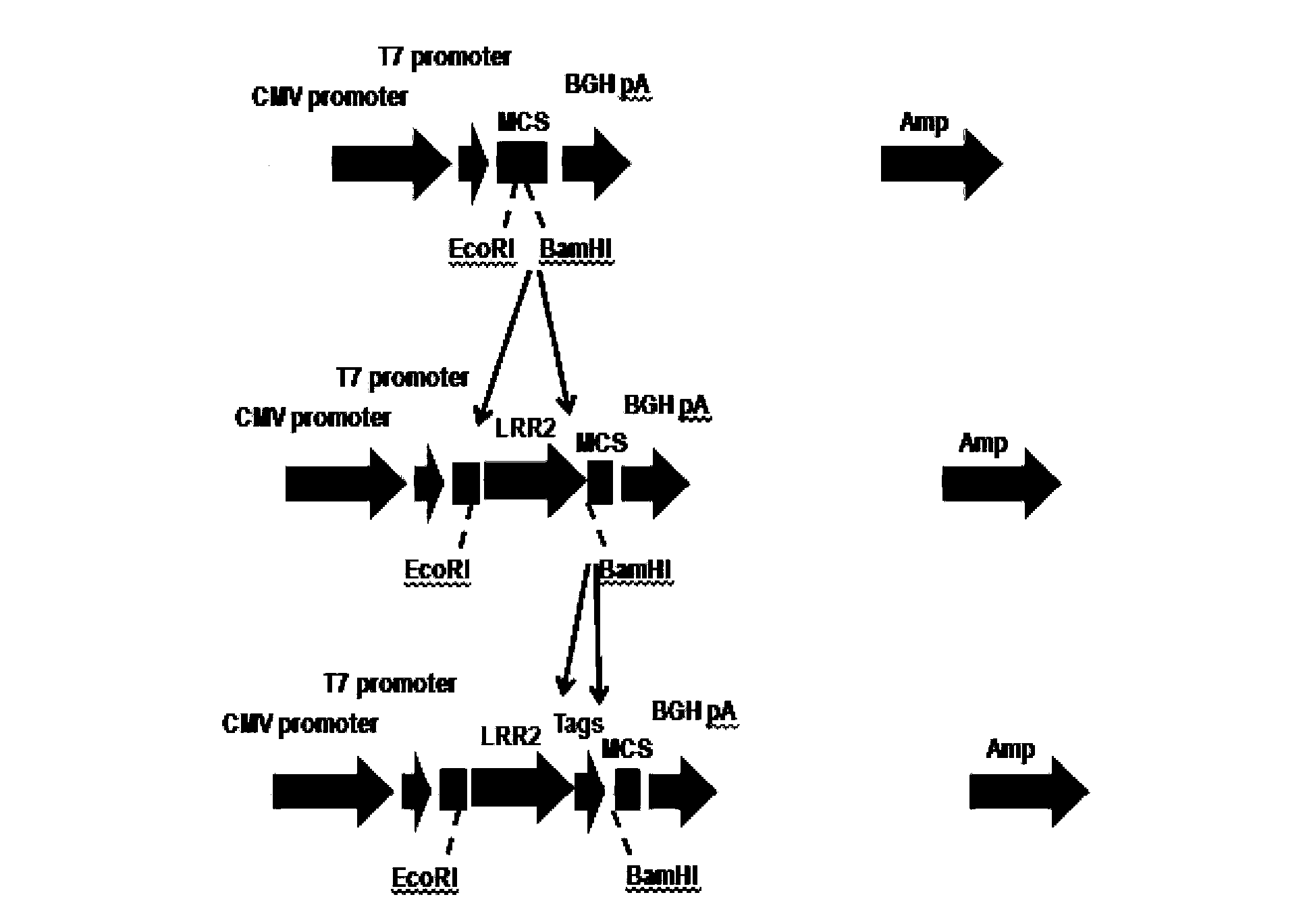
[0128] Example 1 Construction of pcDNA3.1(-) / Slit3LRR2 vector

[0129] Method: The plasmid was constructed by exonuclease method. In the process of constructing the plasmid, a total of three fragments (signal peptide sequence, target sequence, restriction site + tag protein sequence) were amplified by PCR, and the fragments were ligated and inserted three times. The primers used were Primer1, Primer2 and Primer3, respectively.

[0130] Specific steps of exonuclease method:

[0131] 1. Prepare the reaction system: 50ng each of the carrier fragment (recovered from the gel after digestion with BamHI and measure its concentration) and PCR fragment (recovered from the gel after PCR and measure its concentration), 1ul10x exonuclease buffer, supplemented with ddH 2 0 to 10ul;

[0132] 2. Place on ice for 5 minutes, add 1 ul exonuclease III (Takara company) diluted to 20 U / ul, mix well and place on ice for 60 minutes;

[0133] 3. Add 1ul 0.5M EDTA (pH8.0) solution to terminate the r...
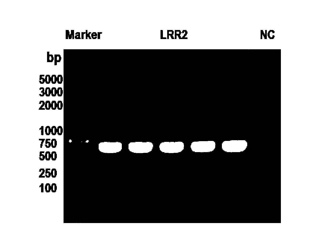
Embodiment 2

[0144] Example 2 Successful Expression of pcDNA3.1(-) / Slit3LRR2

[0145] Method: In order to detect whether the constructed plasmid was successfully expressed, the plasmid was transfected into AD293 cells, and then Western Blot was done with the tag protein 8×His to detect whether the fusion protein was successfully expressed.

[0146] Materials: High glucose DMEM medium and fetal bovine serum were purchased from HyClone Company; transfection reagent Magetran was purchased from Origene Company; AD293 cells were cultured in High glucose DMEM medium containing 10% fetal bovine serum. The inverted microscope is a product of Olympus, and the fluorescent inverted microscope is a product of Nikon.

[0147] step:

[0148] 2.1. Resuscitate and culture the cryopreserved cells, passage 3 to 4 times to make the cells reach a good growth state, and carry out plate detection. Adherent cells were transfected with Magetran reagent.

[0149] 2.2. Inoculate AD293 cells into a 6-well plate a...

Embodiment 3

[0152] The test of embodiment 3 expression amount

[0153] A purified HisGFP protein with a known concentration (20 μg / ml) was used as a control, and the protein expression was determined by Western Blot.

[0154] The specific method is the same as that in Example 2, the cells are first transfected, and then Western Blot detection is performed. The difference is that when running SDS-PAGE electrophoresis, a certain known amount of HisGFP protein is added as a positive control.

[0155] After the exposure results were obtained, the expression level of the protein was calculated by performing grayscale analysis on the bands.


PUM


Abstract

The invention discloses fusion protein containing a leucine-rich repetitive sequence, and a preparation method and application thereof. Concretely, the invention relates to fusion protein, and the fusion protein possesses the following structure from N terminal to C terminal: A-X-E-Y1-Y2 (formula Ia) or A-Y2-Y1-E-X (formula Ib), wherein A is an optional secretion signal peptide, X is a member rich in leucine repetitive sequence 2 (LRRs2) of Slit3 protein (neuronal guidance factor 3), E is a restriction enzyme cutting site, Y1 is a first tag peptide member, Y2 is a second tag short peptide member and the second tag short peptide member is His tag member, and '-' represents a peptide bond or a peptide joint for connecting the above members. The fusion protein is beneficial for correct folding of LRR (leucine-rich repeat) and forming active functional polypeptide, and is capable of simply removing two tag members through once resection enzyme cutting, thereby preparing high-purity high-activity LRR sequence.

Description

technical field [0001] The invention relates to the field of biomolecules, in particular to a fusion protein containing a leucine-rich repeat sequence and a preparation method and application thereof. Background technique [0002] Nerve guiding factor Slit is a secreted extracellular matrix glycoprotein highly conserved in evolution, and its gene was discovered in 1988 to play a role in the formation of the central nervous system. Slit is a group of extracellular secreted proteins with a relative molecular mass of 170-190kDa, and is one of the members of the axon orientation molecular family that plays a guiding role in axon growth and neuron migration. [0003] Slit protein is a multifunctional guiding molecule as a ligand of Roundabout (Robo). It not only has the functions of regulating the growth direction of nerve axons, guiding the migration of nerve cells, and affecting the morphological differentiation of nerve cells. Recent studies have found that Slit also plays a ...

Claims


Application Information

Patent Timeline
IPC IPC(8): C07K19/00C12N15/62C12N15/63C12N5/10C12P21/02
Inventor 李华顺
Owner 李华顺
Why Eureka
Try Eureka