
Method for detecting golden glucose coccus by using immune optical-fiber evanescent-wave biosensor

A biosensor, glucococcus technology, applied in the field of inspection and quarantine, can solve problems such as the inability to form an evanescent wave conduction environment

Inactive Publication Date: 2014-11-05
吉林出入境检验检疫局检验检疫技术中心
View PDF9 Cites 5 Cited by
  • Summary
  • Abstract
  • Description
  • Claims
  • Application Information

AI Technical Summary

Problems solved by technology

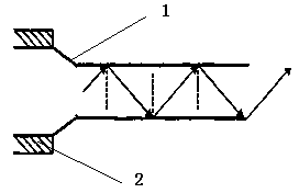
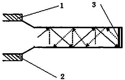
Since the inner diameter of the optical fiber itself can generally reach 200 μm, part of the fluorescence is bound to be emitted from the optical fiber during the fluorescence coupling process. The larger the cone length, the closer the incident angle is to the critical angle, but there are still some rays at the end whose incident angle is smaller than the critical angle. angle, so the propagating light is transmitted into the solution, and an absolute evanescent wave conduction environment cannot be formed.

Method used


Image

  • Method for detecting golden glucose coccus by using immune optical-fiber evanescent-wave biosensor
  • Method for detecting golden glucose coccus by using immune optical-fiber evanescent-wave biosensor
  • Method for detecting golden glucose coccus by using immune optical-fiber evanescent-wave biosensor

Examples


Embodiment 1

[0017] See figure 1 , 2 , an immune optical fiber evanescent wave biosensor probe, which includes a cladding portion 11 and an adsorption portion 12, the end section of the adsorption portion 12 is provided with a silver reflective layer 13; the silver reflective layer 13 is welded Or the vacuum coating method is used to fit the cross-section of the end of the adsorption part; the adsorption part 12 is tapered.

Embodiment 2

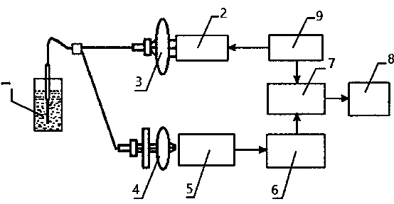
[0019] See Figure 1-3 , an immune optical fiber evanescent wave biosensor, which includes: an optical system, a photoelectric conversion and signal amplification system, and a data acquisition and processing device;

[0020] The optical system includes: a drive circuit 9, a laser 2, a convex lens 3, an optical fiber probe 1, and a filter 4;

[0021] The photoelectric conversion and signal amplification system includes: photomultiplier tube 5 and signal amplifier 6;

[0022] The data acquisition and processing system includes: data acquisition card 7 and laboratory virtual instrument engineering platform (LabVIEW) 8;

[0023] The laser 2 emits laser light through the convex lens 3 to the fiber optic probe, and the fluorescent signal returns; the fluorescent signal is converted and amplified by the photomultiplier tube 5 and the signal amplifier 6; the data acquisition card 7 and the laboratory virtual instrument engineering platform 8 collect, process, and detect signal stren...

Embodiment 3

[0027] 1) Preparation of immuno-optical fiber probe: After peeling the fiber probe, soak it in sodium hydroxide for 10 min, and then soak it in hydrochloric acid for 10 min. After rinsing with deionized water three times, let it dry naturally. The optical fiber was then placed in 10% acetone solution for 1 h, and rinsed with acetone 6 times to remove the residual acetone solution. The surface of the fiber core is activated by reagents such as triaminopropyltriethoxysilane and glutaraldehyde. The treated optical fiber was inserted into the solution of Staphylococcus aureus monoclonal antibody and soaked for 24 hours, and then it was taken out and washed with deionized water to obtain an immune optical fiber probe coated with the captured antigen.

[0028] 2) Detection of Staphylococcus aureus

[0029] Using an immune optical fiber evanescent wave biosensor system described in Example 2, the CdTe-polyclonal antibody conjugate is made into an aqueous solution, the capture a...


PUM


Abstract

The invention discloses a method for detecting golden glucose coccus by using an immune optical-fiber evanescent-wave biosensor. The method comprises the following steps: (1) gluing a silver reflecting layer and coating a golden glucose coccus antibody on an optical-fiber probe of an immune optical-fiber evanescent-wave biosensor; (2) preparing a CdTe-polyclonal antibody conjugate, and making an aqueous solution; (3) inserting the optical-fiber probe capturing the antibody into a solution to be detected, then putting the optical-fiber probe capturing the antibody into the aqueous solution made from the CdTe-polyclonal antibody conjugate, and using the optical-fiber evanescent-wave biosensor for detecting. A 530nm semiconductor laser device is adopted, and a convex lens is arranged at the laser transmitting end of the laser device. The method solves the problem that when a terminal incidence angle of parts of light is smaller than a critical angle, the light is transmitted into the solution, and a fluorescent signal is lost. The detection limit and detection sensitivity of an optical-fiber biosensor system are improved, and the limit of detection of the golden glucose coccus is increased to 5ng / mL.

Description

technical field [0001] The invention belongs to the technical field of inspection and quarantine, and in particular relates to a method for detecting Staphylococcus aureus by an immune optical fiber evanescent wave biosensor. Background technique [0002] The optical fiber evanescent wave biosensor excites the fluorescent signal through the evanescent wave field. When light propagates in an optical fiber with total reflection, it can still generate waves that partially penetrate the interface, and the wave amplitude decays exponentially with the increase of the distance from the interface, so it only exists in a thin layer near the interface, called evanescent wave . Fluorescent immune fiber optic biosensors based on evanescent wave field excitation are a kind of fluorescent biosensors. It mainly combines the characteristics of the evanescent wave field generated when light propagates in the optical fiber and the high specificity and high sensitivity of the antibody-antige...

Claims


Application Information

Patent Timeline
Patent Type & Authority Applications(China)
IPC IPC(8): G01N33/569G01N33/542G01N21/64
CPCG01N33/56938G01N33/54373
Inventor 刘韬刘金华孟日增宋战昀王玮琳聂丹丹刘阳
Owner 吉林出入境检验检疫局检验检疫技术中心
Learn More