Medicine balloon and preparation method thereof

A drug and balloon technology, applied in the field of medical devices, can solve the problems of vascular embolism, inability to treat diseases, and ineffectiveness, and achieve the effects of increasing drug absorption, reducing drug loss, and low systemic toxicity

Active Publication Date: 2015-05-20
SHANGHAI VASOLUTIONS MEDTECH CO LTD
View PDF5 Cites 22 Cited by
  • Summary
  • Abstract
  • Description
  • Claims
  • Application Information

AI Technical Summary

Problems solved by technology

The disadvantages of this invention are: (1) the water-soluble material in the inner layer is tightly wrapped by the degradable polymer in the outer layer, and it is difficult for water molecules to cross the barrier of the outer layer. In many cases, even if soaked for a long time, the water-soluble material in the inner layer cannot On the other hand, even if the water-soluble material in the inner layer is dissolved, its aqueous solution is difficult to completely flow out with the water molecules. Although the solution has certain lubricity in the presence of water, once the coating is dry, The balloon is also adhered to the outer layer; (2) Even if the inner layer is completely removed through soaking treatment, there is no physical connection between the balloon

Method used


Image

  • Medicine balloon and preparation method thereof
  • Medicine balloon and preparation method thereof
  • Medicine balloon and preparation method thereof

Examples


Embodiment 1

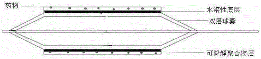
[0033] 1. Double-layer Balloon Preparation

[0034] A double-layer balloon was prepared according to the method described in Chinese Invention Patent Application No. 200610025200.8.

[0035] 2. Coating Preparation

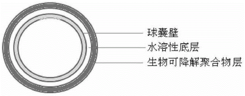
[0036] Weigh 2 g of polyvinylpyrrolidone (PVP, molecular weight 10,000), add it into 5 ml of isopropanol (IPA) solution, and stir well until the PVP is completely dissolved. The inner layer of the double-layer balloon was expanded, immersed in the above solution, and then dried in an oven at 40°C to form a water-soluble bottom layer. The outer diameter of the middle part of the balloon was 30.109mm as measured by a caliper.

[0037] Weigh 2g of poly(lactic-co-glycolic acid) (PLGA) (50 / 50), 0.01g of cyanoacrylate and 300mg of paclitaxel and dissolve in 10ml of tetrahydrofuran (THF) solution, stir well until all components are completely dissolved. Then the balloon coated with the water-soluble bottom layer was immersed in the above solution, dried in an oven at 40...

Embodiment 2

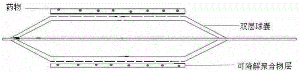
[0040] 1. Double-layer Balloon Preparation

[0041] According to the method described in Chinese Invention Patent Application No. 200610025200.8, a double-layer balloon was prepared.

[0042] 2. Coating Preparation

[0043] Weigh 2g of PVP (molecular weight: 10,000), add it into 5ml of IPA solution, and stir until the PVP is completely dissolved. The inner layer of the double-layer balloon was expanded, immersed in the above solution, and then dried in an oven at 40°C to form a water-soluble bottom layer. The outer diameter of the middle part of the balloon was 30.109mm as measured by a caliper.

[0044] Weigh 2g of PLGA (50 / 50), dissolve 0.01g of cyanoacrylate in 10ml of THF solution, stir well until all components are completely dissolved. Continue to immerse the balloon coated with the water-soluble bottom layer into the above solution, and then dry it in an oven at 40° C., repeat the immersion twice, and dry completely. Finally, a layer of paclitaxel was sprayed on the ...

Embodiment 3

[0047] Take an isolated porcine arterial segment with a diameter similar to that of the balloon, immediately immerse it in pig blood with anticoagulant added, keep a constant temperature of 37°C, pass the drug balloon through a 6F catheter in a folded state and keep it for 1 minute, and then the balloon enters the blood vessel In the inner cavity, the inner balloon is first expanded (12 atm) to achieve the purpose of angiogenesis, and then the outer balloon is injected with normal saline, and the water-soluble bottom layer is dissolved (about 1 minute) and the balloon is withdrawn to form a degradable polymer layer. A thin film adheres to the inner wall of the blood vessel.

[0048] Take another latex tube with a diameter similar to the balloon, and immerse it in 37°C constant temperature phosphate buffer solution (PBS, pH=7.4). ), then inject physiological saline into the outer balloon, and withdraw the balloon after the water-soluble bottom layer dissolves (about 1 minute). ...


PUM

PropertyMeasurementUnit
Thicknessaaaaaaaaaa
Thicknessaaaaaaaaaa
Thicknessaaaaaaaaaa

Abstract

A drug-eluting balloon and a preparation method therefor. The drug-eluting balloon comprises a double-layer balloon, the outer surface whereof is sequentially coated with a water-soluble substrate and a biodegradable polymer layer containing a medicine. During the process of balloon dilatation, physiological saline and related solutions exude through the balloon wall and dissolve the water-soluble substrate, thereby controlling release of the degradable polymer layer containing the medicine.

Description

technical field [0001] The invention belongs to the field of medical devices, and in particular relates to a drug balloon and a preparation method thereof. Background technique [0002] Vascular stenosis is the main cause of morbidity and mortality, and the most commonly used treatment is interventional therapy with drug-eluting stents (DES). DES is a drug mixed with a polymer matrix and coated on the surface of the stent to form a local drug slow release system. After the stent is implanted in the blood vessel, the drug will be slowly released for several weeks to several months. With the support of the stent, it can effectively treat the stenosis or occlusion of the blood vessel. It is currently one of the most commonly used and most effective interventional devices. However, DES also has many disadvantages. For example, the polymer will produce local chronic inflammation; the existence of the stent requires long-term antiplatelet drug treatment; the drug coating is uneve...

Claims


Application Information

Patent Timeline
IPC IPC(8): A61L31/14A61L31/16A61L31/10A61M29/02
CPCA61L29/16A61L29/085A61L29/148
Inventor 张鹏张琳琳郭芳王长松史增佐李中华罗七一
Owner SHANGHAI VASOLUTIONS MEDTECH CO LTD
Why Eureka
Try Eureka