Myoelectricity detection device combined with laparoscope and operating method thereof

A detection device and laparoscopic technology are applied in the field of medical supplies for digestive tract detection, which can solve the problems of large destructive and irreversible effects on the intestinal wall, inability to obtain experimental results, large interference waves in electromyography, and the like, and achieve good economic benefits. and social benefits, reducing interference factors, destructive and less disruptive effects

Active Publication Date: 2016-09-21
THE THIRD AFFILIATED HOSPITAL OF XINXIANG MEDICAL UNIV
View PDF9 Cites 2 Cited by
  • Summary
  • Abstract
  • Description
  • Claims
  • Application Information

AI Technical Summary

Problems solved by technology

[0003] Common electrodes for gastrointestinal myoelectricity mostly use tin electrodes. The material of tin electrodes is soft, not hard enough, and has a low melting point. It is difficult to weld after disconnection.
In addition, the electrodes are usually covered with insulating tubes such as polyvinyl chloride or connected with auxiliary tools such as medical suture needles for guidance. The insulating tubes and medical suture needles will have a destructive and irreversible impact on the intestinal wall, resulting in abnormalities on the EMG. Larger interference waves appear, so ideal experimental results cannot be obtained

Method used


Image

  • Myoelectricity detection device combined with laparoscope and operating method thereof
  • Myoelectricity detection device combined with laparoscope and operating method thereof
  • Myoelectricity detection device combined with laparoscope and operating method thereof

Examples


Embodiment

[0022] The test subject of this example is a rabbit.

[0023] Preparation of epoxy resin / graphene / nano-copper composite electrode

[0024] 1. Preparation of graphene oxide sol: The traditional Hummers method is used to prepare graphene oxide, the graphene oxide is mixed with distilled water, and then the graphene oxide sol is ultrasonically obtained. The concentration of graphene oxide in the graphene oxide sol is 0.01g / mL;

[0025] 2. Graphene oxide supported nano copper powder: CuSO 4 ·5H 2 O was added to distilled water and sonicated until it was completely dissolved to obtain a copper sulfate aqueous solution. Pour the graphene oxide sol and copper sulfate aqueous solution prepared in step 1 into a 250 mL three-necked flask, ultrasonically shake at room temperature for 1 hour, and then place it in a 70°C water bath at stirring speed Under the condition of 300r / min, add hydrazine hydrate aqueous solution and sodium hydroxide aqueous solution successively, continue to stir in a wat...


PUM


Abstract

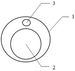
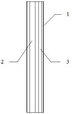
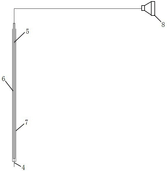
The invention discloses a myoelectricity detection device combined with a laparoscope and an operating method thereof. The myoelectricity detection device comprises a working mirror tube, the laparoscope and a myoelectricity detection electrode. The interior of the working mirror tube is provided with an imaging channel and an electrode channel, both of which are arranged in parallel. The laparoscope is arranged in the imaging channel. The myoelectricity detection electrode is arranged in the electrode channel and comprises a medical cannula needle head, an electrode and an inner cannula and an outer cannula. The inner cannula and the outer cannula differ in internal diameters and are sleeved with each other. The medical cannula needle head is fixed at one end of the outer cannula. The electrode is fixed in the inner cannula. An electrode on one side of the inner cannula is guided into the medical cannula needle head and the front end of the electrode is aligned with the needle point end of the medical cannula needle head. The tail end of an electrode of the inner cannula is connected with a biology signal acquisition system through a wire. The myoelectricity detection device combined with the laparoscope and the operating method thereof have following beneficial effects: an operation wound is small; patient's pain can be effectively mitigated; postoperative recovery is quick; and interference factors caused by an operation are reduced such that an experiment result is more convincing.

Description

Technical field [0001] The invention belongs to the technical field of medical supplies for gastrointestinal detection, and specifically relates to an electromyography detection device combined with a laparoscope and an operation method thereof. Background technique [0002] Colon myoelectric action potentials are generated on slow waves, which are consistent with smooth muscle contraction and are the main driving force of propulsive exercise. Therefore, colonic myoelectric activity, as an objective electrophysiological indicator, can indirectly reflect the colonic dynamics. Slow wave is a relatively regular periodic electrical activity, which controls the contraction rhythm of the intestine, whether it is contracted or not. At present, the recording of myoelectric activity is one of the important methods for colonic motility research. In some tests, because the normal intestinal mucosa contains many blood vessels, it looks red under the microscope. It is not easy to distinguish...

Claims


Application Information

Patent Timeline
Patent Type & Authority Applications(China)
IPC IPC(8): A61B5/0488A61B5/0492A61B1/313A61B1/31A61B1/012A61B5/296
CPCA61B1/012A61B1/31A61B1/3132A61B5/4255A61B5/6848A61B5/6852A61B5/6873A61B2562/0209A61B2562/125A61B5/296A61B5/389
Inventor 杜爱林成竹影姜洪波张玉姣严静路承彪王昊阳张越李文杰代玄张璐
Owner THE THIRD AFFILIATED HOSPITAL OF XINXIANG MEDICAL UNIV
Why Eureka
Try Eureka