Preparation method of bionic artificial bone scaffold with micro-structure

A technology of artificial bone and microstructure, which is applied in the field of biomedical tissue engineering, can solve the problems that the connectivity of the internal pores of the bone scaffold, the diversity of micropores and the characteristics of the micropore structure cannot be well guaranteed, and the preparation time can be shortened and the pain can be relieved. , to ensure the effect of uniformity and diversity

Inactive Publication Date: 2016-10-26
苏州博恩生物科技有限公司
View PDF7 Cites 7 Cited by
  • Summary
  • Abstract
  • Description
  • Claims
  • Application Information

AI Technical Summary

Problems solved by technology

[0010] In order to overcome the requirements of the prior art artificial bone scaffolds that can only guarantee the porosity required by the bone scaffolds, but cannot well ensure the connectivity of the internal pores of the bone scaffolds, the diversity of micropores, and the structural characteristics of micropores, the present invention proposes a A method for preparing an artificial bone scaffold for three-dimensional reconstruction of two-dimensional bone slices. The method applies a dimensionality reduction method: the human body bone as a three-dimensional entity is scanned by CT to obtain a two-dimensional image of the bone slice, and then the two-dimensional Design bone microholes on the pictures, and finally superimpose and reconstruct the pictures to generate a 3D solid model

Method used


Image

  • Preparation method of bionic artificial bone scaffold with micro-structure
  • Preparation method of bionic artificial bone scaffold with micro-structure
  • Preparation method of bionic artificial bone scaffold with micro-structure

Examples


Embodiment Construction

[0033] Specific implementation examples

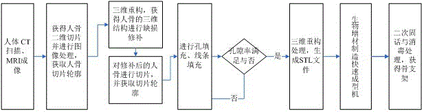
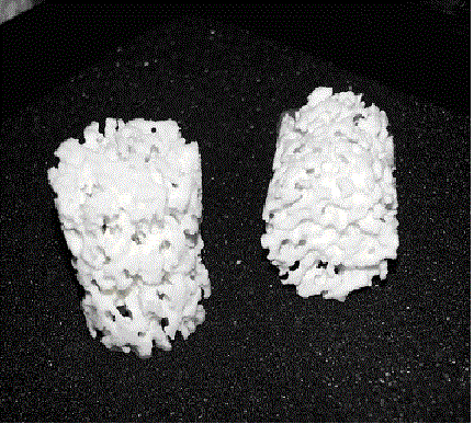
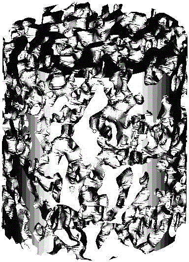
[0034] In this example, a simple cylindrical bone scaffold (diameter d=2.5mm, height h=5mm) is used to describe a preparation method of an artificial bone scaffold based on internal microstructure, which specifically includes the following steps:

[0035] Step 1. Carry out CT or MRI scanning on the replaced bone, and obtain a set of tomographic images of the bone with a total of N layers from bottom to top, where the layer spacing is =0.5mm, the first The area of ​​the cross-sectional figure is . When the pore diameter of the through pores inside the material is 10-40 μm, it allows the growth of fibrous tissue; when the pore diameter is 50-100 μm, it allows the growth of non-mineralized bone-like tissue; when the pore diameter reaches 150 μm or more, it can allow the growth of bone tissue To provide an ideal place, the pores of 200-400 μm are most conducive to the growth of new bone, so the pore size of the micropores filled by t...


PUM

PropertyMeasurementUnit
diameteraaaaaaaaaa

Abstract

The invention discloses a preparation method of a bionic artificial bone scaffold with a micro-structure. The artificial bone scaffold is prepared as follows: replaced bone is scanned through CT or MRI equipment, a group of cross-sectional images of the bone are acquired and subjected to pretreatment, binaryzation, image segmentation, edge tracking, spline fitting and other steps, and a macrostructure is generated; a solution set is obtained by means of a knowledge base, micropore structures meeting conditions of a patient are extracted, micropores are filled in a profile, and the porosity and the fractal dimension of the profile after filling are same as or similar to those of original diseased bone; an STL file is generated and then output to a three-dimensional printer for rapid forming of the bone scaffold.

Description

technical field [0001] The invention belongs to the technical field of biomedical tissue engineering, and relates to a method for preparing an artificial bone support by imitating the Haversen-Volkmann pipeline structure inside a human bone. The invention is based on image processing, combined with fractal theory and three-dimensional modeling, and proposes a new preparation method based on internal microstructure tissue engineering scaffold. Background technique [0002] As a human organ, there are intricate tiny channels inside the bone. Through these channels, capillaries and nerve tissues extend to various parts of human bones. Capillaries transport oxygen, nutrients and various hormones to bone cells, and take away the metabolic waste produced by bone cells. If there is no such microstructure, the cells inside the human bone will die due to lack of nourishment, and it is impossible for the human bone to grow healthily. [0003] The canals in the human bone are actuall...

Claims


Application Information

Patent Timeline
Patent Type & Authority Applications(China)
IPC IPC(8): A61L27/12A61L27/56G06T11/40G06T17/00G06N5/02G06T7/00
CPCA61L27/12A61L27/56A61L2430/02G06N5/022G06T11/40G06T17/00G06T2200/08G06T2207/10081G06T2207/10088G06T2210/41
Inventor 李欣培侯祎波毛海荣杨明明汪焰恩
Owner 苏州博恩生物科技有限公司
Why Eureka
Try Eureka