Fusion protein used for rapidly detecting type I diabetes

A fusion protein, fusion protein technology, applied in fusion polypeptides, microorganism-based methods, hybrid peptides, etc., can solve the problems of low efficiency, insufficient specificity, single assay, etc.

Inactive Publication Date: 2017-05-10
SHANGHAI LINC BIO SCI
View PDF6 Cites 3 Cited by
  • Summary
  • Abstract
  • Description
  • Claims
  • Application Information

AI Technical Summary

Problems solved by technology

[0005] At present, only multiple autoantibodies such as ICA, IAA, GAD, and IA-2A can be measured in a single way in patients with type 1 diabetes, with many steps, high cost, low efficiency, insufficient specificity, and long time.

Method used


Image

  • Fusion protein used for rapidly detecting type I diabetes
  • Fusion protein used for rapidly detecting type I diabetes
  • Fusion protein used for rapidly detecting type I diabetes

Examples


Embodiment 1

[0017] Example 1: Fusion and expression of genes

[0018] (1) The target DNA sequence is artificially synthesized.

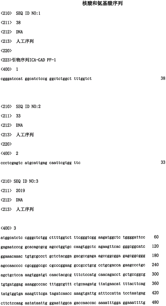
[0019] (2) PCR amplification of the target gene: In order to ensure the fusion expression of the two, Primer5.0 was used to design specific amplification primers according to the sequence of the target gene. The primers were designed with BamH I and Xho I as restriction sites. The primer sequences are:

[0020] cgggatccat ggcatctccg ggctctggct tttggtct SEQ ID NO: 1

[0021] ccctcgagtc atgcattgag caattcgtgg ttc SEQ ID NO: 2

[0022] A high-fidelity DNA amplification system was used to perform PCR cycles using the synthesized DNA sequence as a template to obtain products.

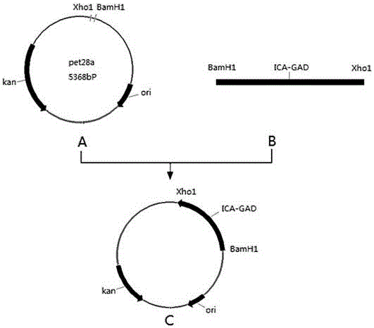
[0023] (3) After amplification, the target gene fragments were recovered according to the AxyPrep DNA Gel Recovery Kit. Insert the target gene into the vector: connect the cDNA product to the vector, transform it into the PET-28a vector, screen according to the marker of the recombinant vec...

Embodiment 2

[0025] Example 2: Protein Purification

[0026] The induced bacteria containing the target protein were collected by centrifugation, ultrasonically disrupted, and the supernatant was collected by centrifugation. In order to improve the purification purity of the target protein, we improved the traditional method of purifying his fusion protein by adding β-mercaptoethanol (to make the final concentration 5mM) in the E. coli supernatant after induction of expression. Then use the imidazole buffer containing 300mM Nacl, 20mM Tris-Hcl and 40mM to bind the target protein to the HIS Trap FF affinity column, and then use the imidazole buffer containing 300mM Nacl, 20mM Tris-Hcl and 500mM to elute the target protein. It was dialyzed overnight at 4°C in PBS buffer, and the protein concentration was determined to be 1 mg / ml by BCA method.

Embodiment 3

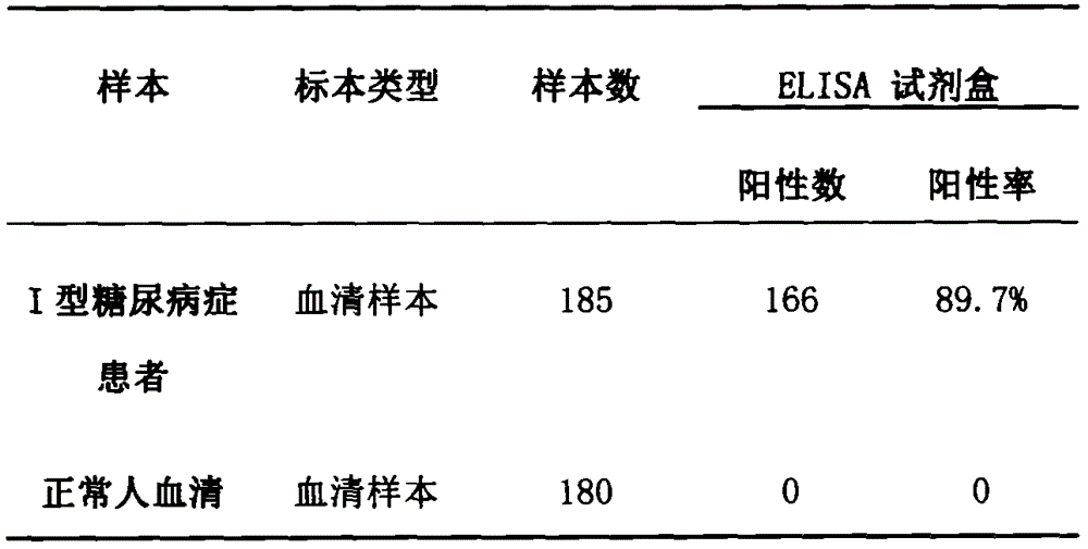
[0027] Example 3: Immunological activity was determined by Elisa detection.

[0028] 1. Coating: Dilute the fusion protein ICA-GAD with 0.05M PH9, carbonate coating buffer to a protein content of 1-10 μg / ml, add 0.1ml to the reaction well of each polystyrene plate, 4 ℃ overnight. The next day, the solution in the well was discarded, and washed 3 times with washing buffer, 3 minutes each time.

[0029] 2. Adding samples: Add 0.1ml of a certain dilution of the sample to be tested to the above-mentioned coated reaction wells, and incubate at 37°C for 1 hour. Then wash. (Make blank wells, negative control wells and positive control wells at the same time).

[0030] 3. Add enzyme-labeled antibody: Add 0.1ml of freshly diluted enzyme-labeled antibody (diluted after titration) to each reaction well, incubate at 37°C for 0.5-1 hour, and wash.

[0031] 4. Add substrate solution for color development: add 0.1 ml of temporarily prepared TMB (tetramethylbenzidine) substrate solution t...


PUM


Abstract

The invention discloses a fusion protein used for rapidly detecting type I diabetes. The fusion protein is a fusion protein ICA-GAD obtained by performing fusion expression on islet cell autoantigen (ICA69) and glutamate decarboxylase (GAD2) genes. The method disclosed by the invention comprises the following steps: performing B cell antigenic epitope analysis on the islet cell autoantigen (ICA69) and glutamate decarboxylase (GAD2) genes through DNA star software, determining the gene sequence, artificially synthesizing target genes (ICA-GAD), inserting the target genes into a protein expression vector pet28a, transforming into BL21(DE3) host bacteria, and adding IPTG to perform inducible expression on a target protein; purifying and separating the expression protein by using a Ni column and a dialysis bag; detecting the activity of the ICA-GAD fusion protein. According to the fusion protein disclosed by the invention, any one or two in antibodies can be detected at a time, the diagnostic efficiency of the type I diabetes is obviously improved, the detection time is shortened, and the hospitalization costs can be reduced.

Description

technical field [0001] The invention relates to a fusion protein for rapid detection of type I diabetes, in particular to the technical field of ICA-GAD fusion protein fused and expressed by ICA69 and GAD2. Background technique [0002] At present, with the improvement of people's living standards, people's sugar intake is high, coupled with less exercise, leading to some people's obesity-induced diabetes. [0003] Diabetes mellitus is a common endocrine and metabolic disease characterized by hyperglycemia. It is caused by hyperglycemia due to insulin secretion defects or impaired biological performance, resulting in glycosuria, which in turn causes fat and protein metabolism disorders, leading to various tissues, especially hyperglycemia. It is chronic damage and dysfunction of eyes, kidneys, heart, blood vessels, and nerves. Diabetes is divided into four types, namely type I diabetes, type II diabetes, gestational diabetes and other special types of diabetes. [0004] Du...

Claims


Application Information

Patent Timeline
Patent Type & Authority Applications(China)
IPC IPC(8): C07K19/00C12N15/70G01N33/68C12R1/19
CPCC07K14/4713C07K2319/00C12N9/88C12N15/70C12N2800/101C12Y401/01015G01N33/6893G01N2800/042
Inventor 罗朝领冯奇马贵芳杜庆辉茅柳娟
Owner SHANGHAI LINC BIO SCI
Why Eureka
Try Eureka