
Nano-fibers with drug two-phase pulse releasing function and preparation method
A technology of nanofiber and pulse release, which is applied in the field of materials science, can solve the problems of poor two-phase pulse control release effect of nanofiber, and achieve the effect of clear parallel structure, simple preparation process and smooth fiber surface
- Summary
- Abstract
- Description
- Claims
- Application Information
AI Technical Summary
Problems solved by technology
Method used
Image

Examples
Embodiment 1
[0030] Example 1: Parallel electrospinning process implementation and preparation of Jonas nanofibers
[0031] Dissolve 1 gram of drug ibuprofen and 7 grams of polyvinylpyrrolidone K60 in 100 ml of absolute ethanol to prepare a side working fluid.
[0032] Dissolve 1 gram of drug ibuprofen and 13 grams of fiber-forming polymer Eudragit L-100 in 100 ml of absolute ethanol to prepare the working fluid on the other side of the side-by-side electrospinning.
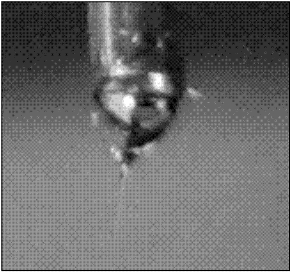
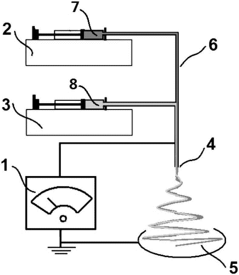
[0033] Put the above two working fluids into the corresponding syringes 7 and 8 respectively, install them on the respective syringe pumps 2 and 3, and connect them to the inlet of the parallel spinning head 4, and connect the high-pressure spinning head 4 and the high-pressure generator 1.
[0034] The speed at which the parallel solution is injected into the parallel spinning head is controlled respectively by two syringe pumps 2 and 3, the parallel flow rate is 1.0mL / h, the distance between the fiber receiving plate 5 and...
Embodiment 2
[0037] Example 2: Structural and Morphological Characterization of Jonas Nanofibers for Biphasic Pulse Release of Drugs
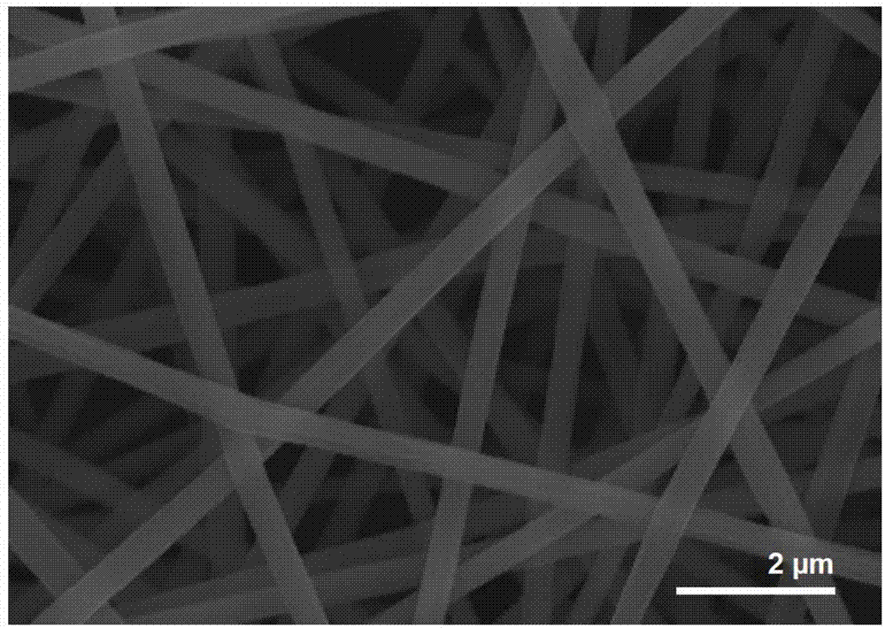
[0038] Field emission scanning electron microscopy (FESEM) was used to observe the surface of the fiber prepared in Example 1 after spraying gold, and the results were as follows image 3 shown. The prepared fiber exhibits a good linear state, no beading structure occurs, the fiber surface is smooth, and the fiber accumulation is uniform. The diameter is 670 ± 110 nm, the distribution is relatively uniform, and the diameter distribution is relatively concentrated.
[0039] The internal structure of the prepared fiber was observed by high-resolution transmission electron microscope (TEM), and the results were as follows: Figure 4 As shown, the bilateral side-by-side structural features of Jonas nanofibers are clear. The internal structure of the fiber is as Figure 5 As shown, the drug 33 is uniformly distributed among the Jonas nanofibers on the left 1...
Embodiment 3
[0040] Example 3: Functional Analysis of Drug Controlled Release of Jonas Nanofibers in Biphasic Pulse Release of Drugs
[0041] According to the 2015 edition of Chinese Pharmacopoeia Appendix ⅩD Release Test Method 2, the RCZ-8A intelligent dissolution tester was used to conduct the in vitro dissolution test on the drug-loaded nanofibers obtained above. The control speed is 50rpm, and the temperature is 37±0.1°C. In the first 2 hours, 900 mL of artificial gastric juice without enzymes was used as the dissolution medium, followed by 900 mL of artificial intestinal juice (pH6.8 phosphate buffer solution) without enzymes as the dissolution medium to investigate the drug release properties of nanofibers in vitro. Sampling 5mL at the scheduled time, filtered through a 0.22 µm microporous membrane to obtain the eluate sample, and immediately replenished with the same volume of isothermal fresh medium. After diluting the sample appropriately, at λ=257 nm, UV-Vis spectrophotometer w...
PUM
| Property | Measurement | Unit |
|---|---|---|
| diameter | aaaaa | aaaaa |
Abstract
Description
Claims
Application Information