ctni-ctnc-ctnt trimer protein and its preparation method and ctni detection kit

A ctni-ctnc-ctnt, trimer technology, applied in the field of time-resolved fluorescence quantitative detection kits, can solve the problems of protein insolubility and difficulty, and achieve the effects of stable labeled products, reliable results and high sensitivity

Active Publication Date: 2021-04-30
郑忠亮
View PDF2 Cites 0 Cited by
  • Summary
  • Abstract
  • Description
  • Claims
  • Application Information

AI Technical Summary

Problems solved by technology

[0015] Our previous experimental results show that the coupled prokaryotic expression of cTnI and cTnC dimers can only produce inclusion bodies, the protein is insoluble, and there are still difficulties in practical use

Method used


Image

  • ctni-ctnc-ctnt trimer protein and its preparation method and ctni detection kit
  • ctni-ctnc-ctnt trimer protein and its preparation method and ctni detection kit
  • ctni-ctnc-ctnt trimer protein and its preparation method and ctni detection kit

Examples


Embodiment 1

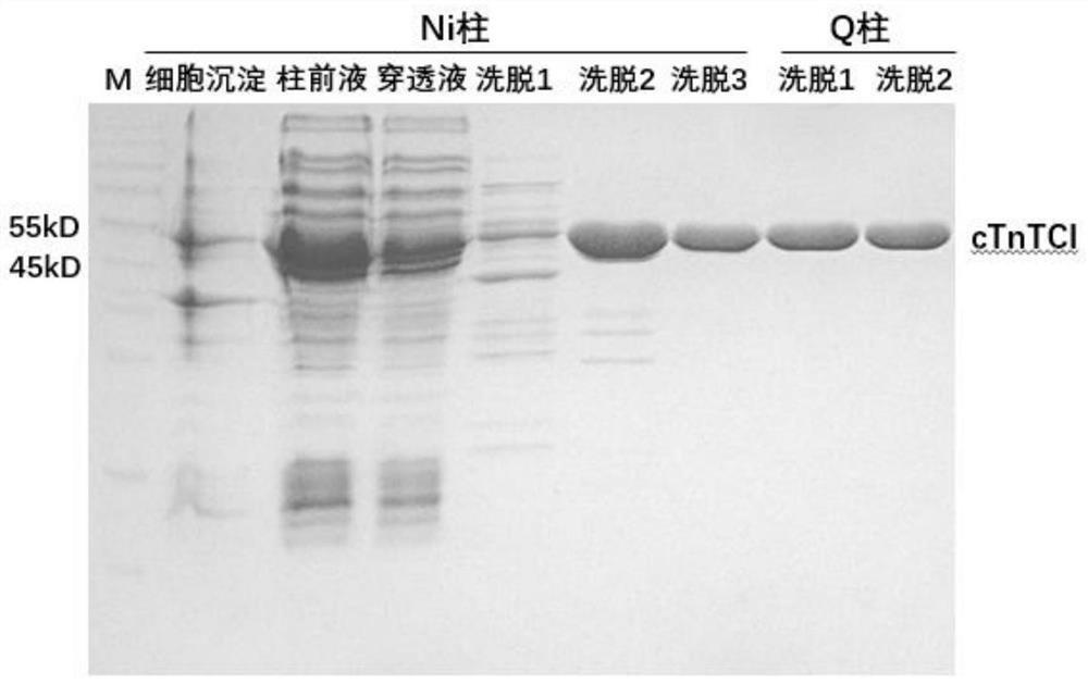
[0051] Embodiment 1: Cloning, expression and purification of cTnI fusion protein

[0052] ①According to the direction from the N-terminal to the C-terminal of the amino acid sequence, artificially synthesize the "cTnI-cTnC-cTnT" triplet gene sequence, as shown in SEQ ID NO.2.

[0053] ② The "cTnI-cTnC-cTnT" gene sequence was cloned into the multiple cloning insertion sites of BamH I and Xhol I of the pET28a plasmid expression vector to obtain the expression plasmid pET28a-cTnI.

[0054] ③ The cloned expression plasmid pET28a-cTnI was transformed into Escherichia coli BL21(DE3), plated and cultured overnight.

[0055] ④ The next morning, pick a single colony and inoculate it into LB medium, and culture it at 37°C until the optical absorption value (OD) of the bacterial solution reaches 1.0, and then add 1 μg / mL IPTG to induce expression. Bacteria were collected by centrifugation after incubation at 37°C for 4 hours.

[0056] ⑤ Add 100mL of 100mM Tris-HCl buffer (pH7.4) to the...

Embodiment 2

[0058] Embodiment 2: ELISA quantitative detection of cTnI active protein

[0059] ①The total protein content of the purified cTnI-cTnC-cTnT trimer protein was measured using the BCA protein content assay kit produced by Thermal Power Company, and the three batches of purified proteins were detected and error analysis was performed. The results are shown in Table 1.

[0060] ②Use the cTnI ELISA kit produced by CloneTech to detect the active protein content of cTnI. The above three batches of purified proteins were tested and analyzed for errors. The results are shown in Table 1.

[0061] Table 1. Determination of total protein content by BCA method and determination of cTnI active protein content by ELISA method

[0062]

Embodiment 3

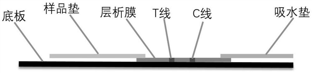
[0063] Embodiment 3: the preparation of immunochromatography kit

[0064] (1) Fluorescent microsphere-labeled antibody:

[0065] ① Take 0.5mL microspheres, centrifuge to get the precipitate, add MES buffer of pH 6.0 to wash twice.

[0066] ②Activation: 114 μL of 50 mM carbodiimide (EDAC) and 114 μL of 50 mM hydroxysulfosuccinimide (Sulfo-NHS) were added to the washed microspheres respectively, and shaken at room temperature for 1 h.

[0067] ③ Centrifuge the microspheres and wash twice with PB buffer.

[0068] ④ Add an appropriate amount of mouse-derived cTnI monoclonal antibody and shake at room temperature for 2 hours.

[0069] ⑤ Centrifuge the microspheres, add 50mM hydroxylamine buffer to quench the reaction, shake at room temperature for 5min; then centrifuge the microspheres, add 50mM hydroxylamine buffer to completely quench the reaction, shake at room temperature for 30min

[0070] ⑥ Centrifuge the microspheres, add blocking solution (0.5% casein, 10mM PB), shake at...


PUM


Abstract

The invention discloses a cTnI-cTnC-cTnT trimer protein, a preparation method and a cTnI detection kit. The trimeric protein is formed by connecting cTnI protein, cTnC protein and cTnT protein in sequence. The preparation method is to artificially synthesize the coding gene of the trimer protein, clone it into an expression vector to obtain an expression plasmid, then transform it into Escherichia coli, and induce expression with IPTG; after the culture is completed, the culture solution is stirred and shaken with a Tris-hydrochloric acid buffer to precipitate bacteria Resuspend, ultrasonically break the wall, centrifuge to take the supernatant, and pass it through a nickel column, a strong anion exchange column Q column and a Sephedex G200 molecular sieve chromatography column in sequence to obtain purified active protein. Using this protein as a positive standard, a time-resolved fluorescence quantitative detection kit with rare earth fluorescent microspheres as a carrier was prepared for the detection of cTnI in samples, which is stable, sensitive, quantitative, simple, rapid and reliable.

Description

technical field [0001] The present invention relates to a cTn trimer protein and its preparation method and cTnI detection kit, in particular to a cTnI, cTnC and cTnT fusion trimer protein of three subunits of troponin and its genetic engineering expression and a detection kit Time-resolved fluorescence quantitative detection kit for three subunit proteins. Background technique [0002] Troponin is composed of three subunits, T, C, and I. Together with tropomyosin, it regulates the interaction between actin and myosin by regulating the activity of calcium ions on striated actin ATPase. After myocardial injury, the cardiac troponin complex is released into the blood, and after 4 to 6 hours, it begins to increase in the blood, and the elevated troponin I can remain in the blood for a long time of 6 to 10 days. [0003] The role of troponin in acute myocardial infarction is huge. Whether it is thrombolysis, percutaneous coronary intervention (PCI), or coronary artery bypass gr...

Claims


Application Information

Patent Timeline
Patent Type & Authority Patents(China)
IPC IPC(8): C07K14/47C12N15/12C12N15/70G01N33/68G01N33/543
CPCC07K14/4716C12N15/70G01N33/54313G01N33/6887G01N2333/4712
Inventor 郑忠亮
Owner 郑忠亮
Why Eureka
Try Eureka