

Application of Corylifol A in preparing anti-radiation drug
An anti-radiation and drug technology, applied in the field of anti-radiation medicine, can solve the problems of weak radiotherapy selectivity, affecting the treatment effect of tumor patients, and treatment failure.
- Summary
- Abstract
- Description
- Claims
- Application Information
AI Technical Summary
Problems solved by technology
Method used
Image

Examples
Embodiment 1
[0028] Example 1 Extraction of Corylifol A and its characterization
[0029] Extraction of Corylifol A
[0030] 15 kg of psoralen fruit, crushed, soaked and extracted with 75% ethanol at room temperature for 3 times, each time for 24 to 36 hours, concentrated under reduced pressure to obtain 5 L of extract, and the extract was applied to silica gel column chromatography, with a volume ratio of 100:1 Gradient elution to 0:100 petroleum ether-ethyl acetate system to obtain 259g of extract at 5:5; the above extract was subjected to silica gel column chromatography, and dichloromethane- Gradient elution with ethyl acetate system, detection by thin layer chromatography, 5 g of fractions containing Corylifol A were collected, and then preparative liquid chromatography was eluted with a methanol-water system with a volume ratio of 75:25 to 95:5 to obtain Pure Corylifol A 1.6g.
[0031] Characterization of Corylifol A
[0032] Pale yellow powder, by electrospray ion mass spectromet...
Embodiment 2
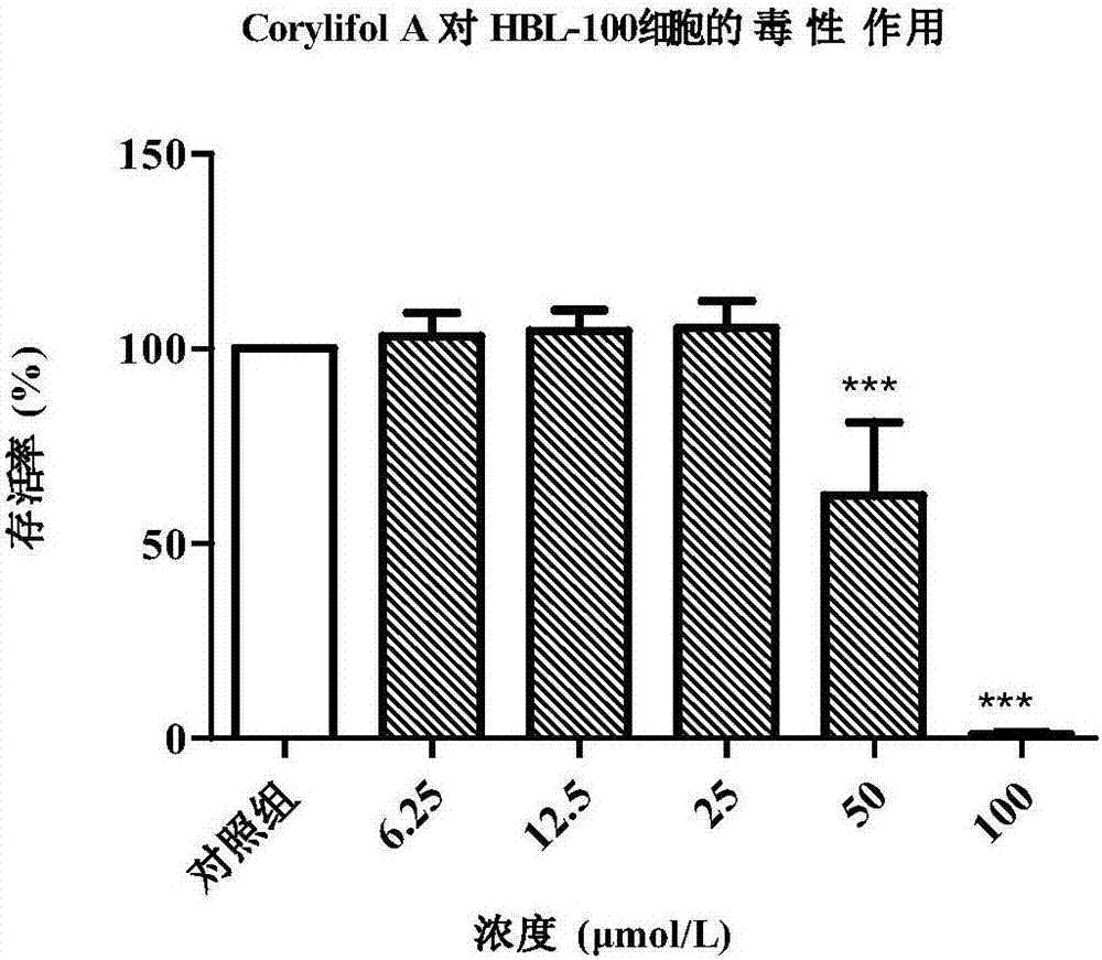
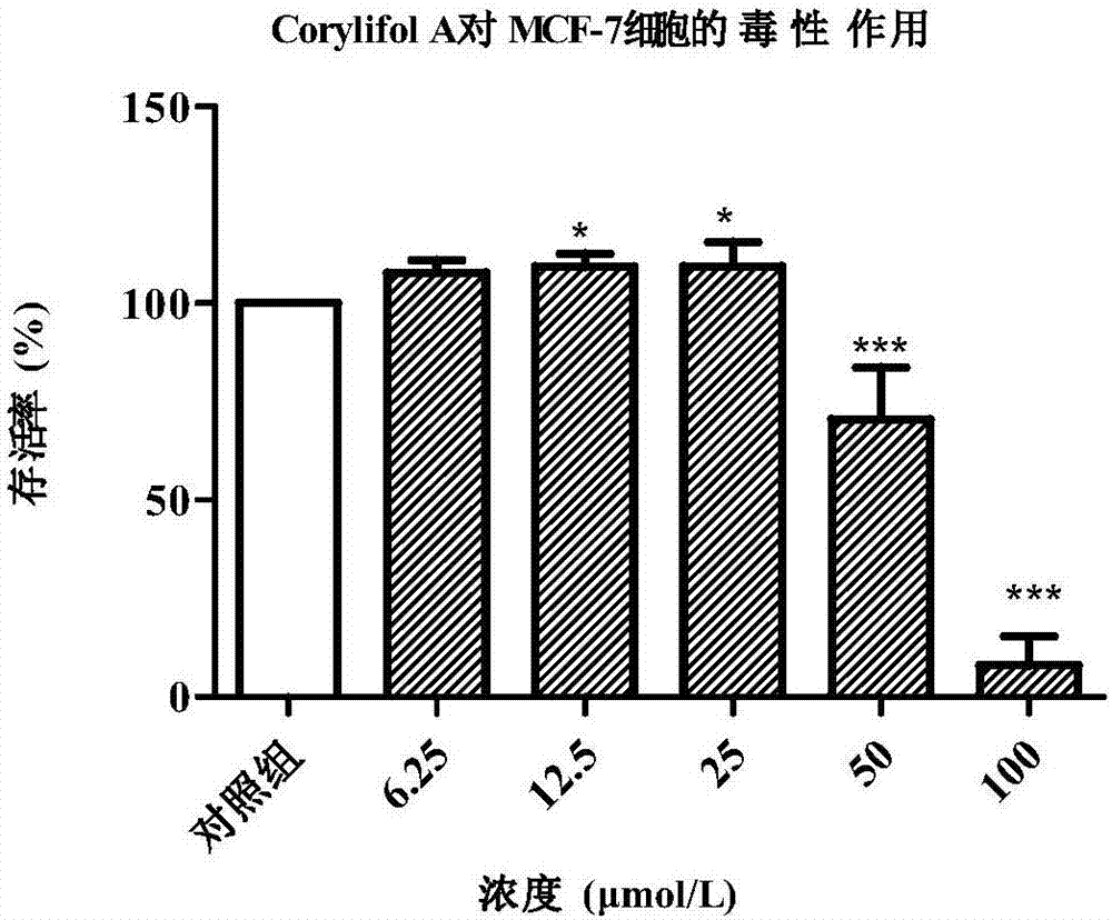
[0034]Example 2 In vitro cytotoxicity test of Corylifol A
[0035] 2.1 Cell culture
[0036] HBL-100 cells (ATCC) in RPMI-1640 medium (containing 10% fetal bovine serum, 100U / mL penicillin and 0.1mg / mL streptomycin), MCF-7 cells (ATCC) in DMEM / High Gluose medium (containing 10% fetal bovine serum, 100 U / mL penicillin and 0.1 mg / mL streptomycin), at 37°C, 5% CO 2 , cultured in an incubator with saturated humidity. When the cells adhere to the wall and grow to a density of 70-80%, they are subcultured at a ratio of 1 / 2. The number of cells in good growth phase was used for experiments.
[0037] 2.2 Cytotoxicity experiment
[0038] CCK-8 method was used to test the effect of test drugs on the viability of HBL-100 and MCF-7 cells.
[0039] (1) Cell inoculation: cells in good logarithmic growth phase were taken, digested with 0.25% trypsin and prepared into cell suspension, and 7×10 3 Cells / well, 100 μL of cell suspension was uniformly inoculated in a 96-well plate (the periph...
Embodiment 3
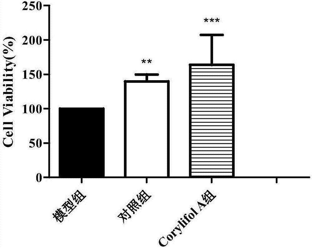
[0047] Example 3 Cell Anti-radiation Experiment of Corylifol A
[0048] 3.1 Cell radiation resistance experiment
[0049] Under 2.2 of Example 2, under the condition that there is no significant difference in cell viability between the corylifol A group and the control group, 25 μmol / L was selected to test its radiation damage repairing effect on HBL-100 and MCF-7 cells. The experiment was divided into blank group (no irradiation, no administration), control group (irradiation, no administration), and corylifol A group (ie, experimental group, irradiation, administration).
[0050] Get the cells with good logarithmic growth phase cultured in 2.1 of Example 2, digest them with 0.25% trypsin and prepare a cell suspension, and prepare 7×10 3 1 / well, 100 μL of cell suspension was uniformly inoculated in a 96-well plate (the periphery of the experimental well was filled with sterile PBS); after culturing in the incubator for 12 h, the 96-well plate was taken out, and 100 μL of the...
PUM
 Abstract
Description
Claims
Application Information