Porcine transmissible gastroenteritis virus (TGEV) fusion protein as well as preparation method and application thereof

A fusion protein and Escherichia coli technology, applied in biochemical equipment and methods, viruses, viral peptides, etc., can solve the problems of low drug intake and ineffective penetration of cell membranes

Active Publication Date: 2018-04-20
WUHAN UNIV
View PDF3 Cites 2 Cited by
  • Summary
  • Abstract
  • Description
  • Claims
  • Application Information

AI Technical Summary

Problems solved by technology

Generally, in the process of transmembrane transduction, biological macromolecules cannot effectively penetrate into cells due to the relevant characteristics of the cell membrane, resulting in low drug intake. The discovery of TAT protein transduction peptide provides a new method to make up for this deficiency. Safe and effective transmembrane transduction method with good application prospects

Method used


Image

  • Porcine transmissible gastroenteritis virus (TGEV) fusion protein as well as preparation method and application thereof
  • Porcine transmissible gastroenteritis virus (TGEV) fusion protein as well as preparation method and application thereof
  • Porcine transmissible gastroenteritis virus (TGEV) fusion protein as well as preparation method and application thereof

Examples


Embodiment 1

[0043] [Example 1] Preparation of fusion gene S1-TAT

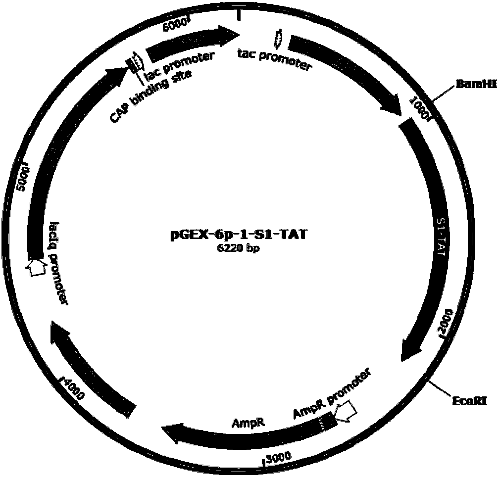
[0044] The S1 gene sequence (KU729220) of TGEV TH-98 strain (KU729220) was obtained from the gene bank of the National Center for Biotechnology (NCBI). The nucleotide sequence connected to TAT at the 3' end is named: S1-TAT, and its sequence is the nucleotide sequence shown in SEQ ID NO.1.

[0045] The recombinant gene S1-TAT was artificially synthesized and connected to the expression vector pGEX-6p-1, that is, the recombinant expression plasmid pGEX-6p-1-S1-TAT was synthesized.

Embodiment 2

[0046] [Example 2] Construction of pGEX-6p-1-S1-TAT engineering bacteria

[0047] Take 1 μl of the recombinant expression plasmid pGEX-6p-1-S1-TAT to transform Escherichia coli E.coli BL21 (DE3) competent cells, and screen out positive transformants through bacterial liquid PCR and gene sequencing identification, and the obtained positive transformants are able to express The recombinant genetic engineering strain E.coli BL21(DE3) (pGEX-6p-1-S1-TAT) of the fusion protein S1-TAT.

[0048] The strain was sent to the China Center for Type Culture Collection on December 12, 2017. It was classified and named: Escherichia coli BL21(DE3)(pGEX-6p-1-S1-TAT)Escherichia coli BL21(DE3)(pGEX-6p -1-S1-TAT), deposit number: CCTCC NO: M 2017785, address: China. Wuhan. Wuhan University.

Embodiment 3

[0049] [Example 3] Expression of Genetic Engineering Fusion Protein S1-TAT

[0050] ① Inoculate a single colony of recombinant genetically engineered strain E.coli BL21(DE3)(pGEX-6p-1-S1-TAT) in 20ml LB liquid medium containing 100μg / ml Amp, and culture overnight at 37°C for 10-12h.

[0051] ②The next day, transfer to 20ml fresh LB liquid medium containing 100μg / ml Amp at a ratio of 1:100, culture at 37°C for about 3 hours until the OD value is about 0.6-0.8, add the inducer IPTG to a final concentration of 0.5mM, induced expression at 250rpm in a shaker at 37°C for 4h.

[0052] ③ Centrifuge the bacterial solution at room temperature for 1-2 minutes, discard the supernatant, resuspend the bacterial cells with an appropriate amount of PBS buffer for the pellet, break with 400W ultrasonication for 3 minutes, break for 3 seconds, stop for 5 seconds, then centrifuge for 2 minutes at room temperature, take 50 μl of the supernatant and mix with The 5×SDS-PAGE loading buffer was mix...


PUM


Abstract

The invention discloses porcine transmissible gastroenteritis virus (TGEV) fusion protein as well as a preparation method and application thereof. The fusion protein S1-TAT provided by the invention comprises a sequence of a main antigenic site of the S protein of the TGEV and 11 core amino acids bound to a basic amino acid-rich region of the TAT protein transduction peptide at the C-terminal of the sequence. Mice are immunized with the fusion protein S1-TAT by means of intraperitoneal injection or intragastric administration, so that the production of specific serum IgG antibodies and sIgA mucosal antibodies can be effectively induced, and good immunogenicity is achieved; TAT can carry S1 protein which is expressed in fusion with the TAT, and passes through intestinal wall cells; the fusion protein S1-TAT has no effect on the growth of the mice. Therefore, the TGEVS1 protein and the TAT are expressed in a fusion way, so that a new method is provided for prevention of TGEV infection, and a foundation is also laid for the development of a novel vaccine for the TGEV.

Description

technical field [0001] The invention belongs to the technical field of genetic bioengineering, and in particular relates to a fusion protein of porcine transmissible gastroenteritis virus S1 and TAT, a preparation method thereof and related applications. Background technique [0002] Porcine transmissible gastroenteritis virus (TGEV), which belongs to the genus Coronaviridae, is a single-stranded positive-sense RNA virus and the main pathogen that causes acute porcine transmissible gastroenteritis disease. The virus mainly infects the small intestine of pig populations, and can rapidly replicate and proliferate, destroying the epithelial cells of the small intestinal mucosa, leading to intestinal dysfunction, intestinal atrophy, and young pigs with symptoms such as vomiting, diarrhea, and dehydration within hours of infection. The virus is explosive in seasons, especially in winter and spring. Generally, the mortality rate can reach 100% within 3-7 days after the onset. Adul...

Claims


Application Information

Patent Timeline
Patent Type & Authority Applications(China)
IPC IPC(8): C07K19/00C12N15/62C12N15/70C12N1/21A61K39/225A61P31/14
CPCA61K39/12A61K2039/552C07K14/005C07K2319/10C12N15/70C12N2770/20022C12N2770/20034
Inventor 徐进平王芳
Owner WUHAN UNIV
Why Eureka
Try Eureka