Cartilage defect repair material and preparation method thereof

A repair material and cartilage technology, applied in the field of medical devices, can solve the problems of immune rejection, slow process of allogeneic tissue reconstruction, disease transmission, etc., and achieve the effects of easy survival, reducing patient trauma, and strong biocompatibility

Inactive Publication Date: 2018-06-01
THE FIRST AFFILIATED HOSPITAL OF ARMY MEDICAL UNIV
View PDF5 Cites 6 Cited by
  • Summary
  • Abstract
  • Description
  • Claims
  • Application Information

AI Technical Summary

Problems solved by technology

The use of allogeneic osteochondral grafts faces the risk of immune rejection and disease transmission, and the process of remodeling of allogeneic tissue in the cartilage defect area is also slow

Method used


Image

  • Cartilage defect repair material and preparation method thereof
  • Cartilage defect repair material and preparation method thereof
  • Cartilage defect repair material and preparation method thereof

Examples


Embodiment 1

[0028] Example 1. Preparation of cartilage microparticle-collagen complex.

[0029] 1. Preparation of cartilage particles:

[0030] The fresh articular cartilage of the donated juvenile human body was cut under sterile conditions to form 1×1×1 mm cartilage particles ( figure 1 ). The survival assay showed that the cell survival rate before transplantation was 80%-95%. DMEM medium can be used for temporary storage, or cryopreservation.

[0031] 2. Preparation of collagen material:

[0032] 200g (wet weight) of articular cartilage from healthy adult pigs was intercepted, freeze-dried at 0-10°C, pulverized, aseptically processed, digested, salted out, and dialyzed (see "Acellular osteochondral scaffold combined with autologous bone marrow mesenchymal stem cells to repair sheep. Research on Osteochondral Defects". Chinese Journal of Joint Surgery (Electronic Edition), 2010, 06: 765-771.) Type Ⅱ collagen 10g was prepared. It can be used after sterility test.

[0033] 3. Prepa...

Embodiment 2

[0036] Trim the porcine cartilage defect to make the residual cartilage edge smooth, stable and free from loosening. The cartilage microparticle-collagen composite prepared in Example 1 was aseptically injected into the cartilage defect area, and the temperature of the implanted area was raised to 37° C. and maintained for 6-10 minutes, and the sol matrix material phase changed into gel. The surgical joint was flexed and extended, and the implantation operation was completed after observing that the graft did not fall off in the cartilage defect area.

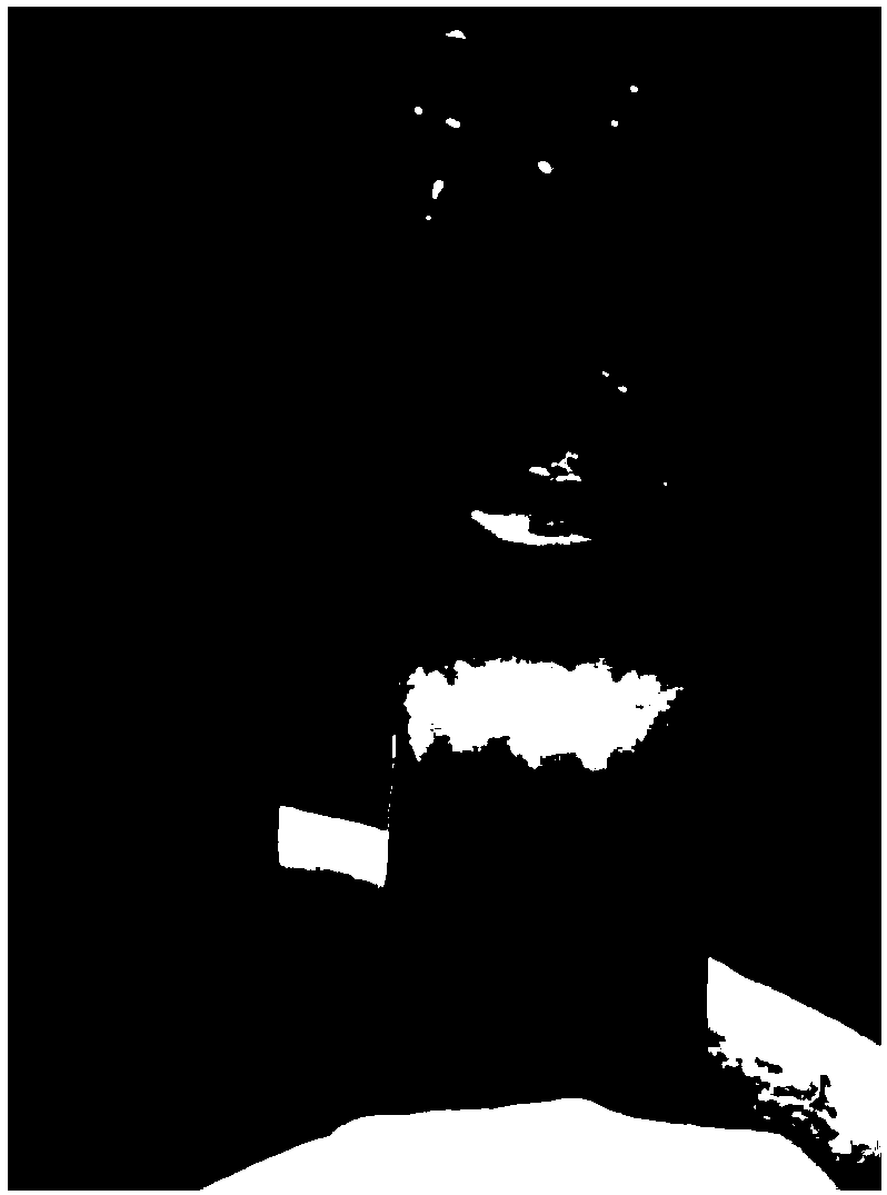
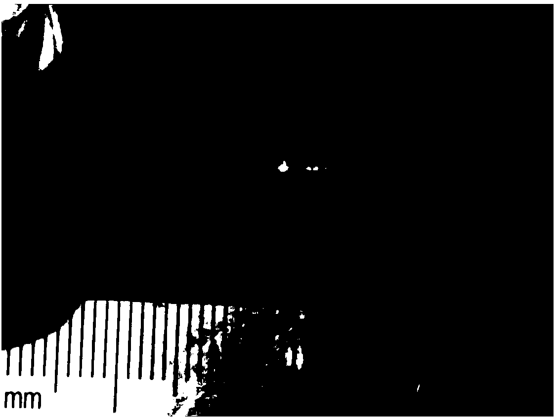
[0037] 3-6 months after surgery, the specimens were taken out for type II collagen immunohistochemical staining analysis. The results are shown in figure 1 and figure 2 .

[0038] The results showed that in the cartilage defect site where the cartilage particle-collagen complex was not injected, the cartilage defect was obvious ( figure 1 ); while the cartilage defect site injected with cartilage particle-collagen complex s...


PUM


Abstract

The invention belongs to the field of medical instruments, and discloses a cartilage defect repair material. The cartilage defect repair material is prepared from cartilage microparticles and II-typecollagen. The cartilage microparticles in the cartilage defect repair material are wide in source, which can be autogenous cartilage microparticles and allogenic cartilage microparticles, so that repeated operations can be avoided, and a wound in a patient is reduced. The cartilage defect repair material disclosed by the invention contains cartilage cells with repair capacity and active extracellular matrixes, and furthermore, a collagen material is high in biocompatibility, so that cells multiplied in the cartilage defect repair material or cells migrating from the body of a host are easy tosurvive and can be further multiplied. The cartilage defect repair material disclosed by the invention is required to be cultivated in vitro, and can be directly applied to repair of a cartilage defect.

Description

technical field [0001] The invention belongs to the field of medical devices, in particular to a cartilage defect repair material and a preparation method thereof, in particular to a cartilage particle-collagen composite repair material and a preparation method thereof. Background technique [0002] Cartilage is the main scaffolding component of the early embryo and is gradually replaced by bone as the fetus develops, a process that continues into the postnatal period. It is a specialized dense connective organ with firm and elastic intercellular substance. Cartilage tissue is widely distributed in the fetus and young, and is gradually replaced by bone tissue. In adults, only some cartilages are scattered, and their functions vary depending on the location. For example, the articular cartilage in the articular surface of the bone plays an important role in the human body, such as bearing load and reducing the friction between the bones between the joints. [0003] Articul...

Claims


Application Information

Patent Timeline
Patent Type & Authority Applications(China)
IPC IPC(8): A61L27/24A61L27/36
CPCA61L27/24A61L27/3612A61L27/3654A61L2430/06
Inventor 段小军杨柳王富友
Owner THE FIRST AFFILIATED HOSPITAL OF ARMY MEDICAL UNIV
Why Eureka
Try Eureka