Cystatin C fluorescent microsphere immunochromatographic quantitative test strip, and test card thereof

A fluorescent microsphere, quantitative detection technology, applied in the field of immunodetection, to reduce non-specific binding, improve performance, clear detection lines and quality control lines

Inactive Publication Date: 2018-08-14
河南省生物工程技术研究中心
View PDF6 Cites 12 Cited by
  • Summary
  • Abstract
  • Description
  • Claims
  • Application Information

AI Technical Summary

Problems solved by technology

However, for cystatin C, the relevant techniques and detection tools for its quan...

Method used


Image

  • Cystatin C fluorescent microsphere immunochromatographic quantitative test strip, and test card thereof
  • Cystatin C fluorescent microsphere immunochromatographic quantitative test strip, and test card thereof
  • Cystatin C fluorescent microsphere immunochromatographic quantitative test strip, and test card thereof

Examples


Embodiment 1

[0045] Example 1. Preparation of Cystatin C Monoclonal Antibody Labeled with Fluorescent Microspheres (EDC Method)

[0046] Take one 1.5ml centrifuge tube, mark it well, add 500μl, 0.02M, PH=7.5 Tris-HCl, and then add 10μl carboxyl fluorescent microspheres (lanthanide europium (Eu) with a particle size of 180nm 3+ ) chelate) and 60ul 2mg / ml EDC activator, shake and mix, place on a constant temperature shaker for 20min, centrifuge at 15000r / min for 10min, discard the supernatant, add 1000ul 0.02M, PH=7.5Tris-HCl Redissolve the precipitate; add 40 μg of mouse cystatin C monoclonal antibody, mix well, put it on a constant temperature shaker to oscillate and couple, centrifuge at 15000rpm / min for 10min, remove the supernatant, add 500ul reconstitution solution to redissolve the precipitate, add Block with 50 μl of blocking solution containing 10% BSA for 1 hour, and store at 4°C for later use.

Embodiment 2

[0047] Embodiment 2, the preparation of sample pad

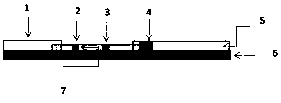
[0048] Take a commercially available glass fiber membrane as the sample pad substrate, and divide it into three groups, 6 in each group:

[0049] The first group: without treatment, spray the fluorescent microsphere-labeled cystatin C monoclonal antibody prepared in Example 1 onto the glass fiber membrane in an amount of 10 ul / ml with a three-dimensional gold spray film apparatus, and heat Dry for 2 hours;

[0050] The second group: use the treatment liquid for soaking treatment, then place it in a constant temperature drying oven at 37°C to dry for 2 hours, and then perform the same spraying operation, wherein the treatment liquid composition is: Tris with a working concentration of 0.05M and a pH of 7.5-8.0 – HCl buffer, BSA at a mass concentration of 0.05-0.5%, and 1% sucrose and 0.05% NaN 3 ;

[0051] The third group: use the treatment liquid for soaking treatment, then place it in a constant temperature drying oven a...

Embodiment 3

[0054] Embodiment 3, the preparation of nitrocellulose membrane (NC membrane)

[0055] Use 0.02M pH7.5 PBS buffer to dilute the rabbit-derived Cystatin C polyclonal antibody to a concentration of 2mg / mL, and use 0.02M pH7.5 PBS buffer to dilute the goat anti-mouse IgG polyclonal antibody to a concentration of 1mg / mL , The resulting diluted solution was streaked on the NC membrane to form a test line (T line) and a quality control line (C line), the two lines were separated by 5mm, dried overnight at 37°C, and stored in a dry environment at room temperature for later use.


PUM

PropertyMeasurementUnit
Particle sizeaaaaaaaaaa

Abstract

The invention relates to a cystatin C fluorescent microsphere immunochromatographic quantitative test strip, and a test card including the test strip. The test strip comprises a base plate, a sample pad, a binding pad, a nitrocellulose membrane and an absorbent paper, the binding pad is provided with a fluorescent microsphere-labeled cystatin C monoclonal antibody, and the nitrocellulose membraneis coated with a detection line formed by a cystatin C polyclonal antibody or a monoclonal antibody and a quality control line formed by a sheep anti-mouse IgG polyclonal antibody. The test strip realizes the rapid and quantitative detection and analysis of cystatin C, has the advantages of high sensitivity, good accuracy, wide linear range, short detection time and convenience in use, and can meet bedside emergency and onsite rapid detection demands.

Description

technical field [0001] The invention belongs to the technical field of immune detection, and in particular relates to a fast and highly sensitive cystatin C fluorescent microsphere immunochromatographic quantitative detection test strip and a test card containing the test strip. Background technique [0002] The gene encoding of Cystatin C (CysC for short) is located on the short arm of human chromosome 20. The Cystatin C gene contains a housekeeping gene, which can be constantly and continuously transcribed and expressed in all tissues. Cystatin C can be synthesized by all nucleated cells and quickly secreted outside the cells, without tissue specificity, it can be distributed in almost all tissues of the body, such as kidney, liver, pancreas, intestine, stomach, lung and placenta, and in cerebrospinal fluid and semen The concentration is the highest in the medium and the lowest in the urine. Cystatin C has a small molecular weight (13KD), is positively charged under physi...

Claims


Application Information

Patent Timeline
IPC IPC(8): G01N33/68G01N33/558
CPCG01N33/558G01N33/68
Inventor 王云龙徐瑞芳李玉林王继创
Owner 河南省生物工程技术研究中心
Why Eureka
Try Eureka