Preparation method and application of autologous fibroblasts
A fibroblast and autologous technology, applied in the field of fibroblasts, can solve the problems of large cell damage, low preparation and separation efficiency, complicated preparation process, etc., and achieves high cell safety, low skin extraction area requirements, and high preparation efficiency. Effect
- Summary
- Abstract
- Description
- Claims
- Application Information
AI Technical Summary
Problems solved by technology
Method used
Image


Examples
Embodiment 1
[0041] Embodiment 1: Human autologous fibroblasts are isolated and expanded and cultivated
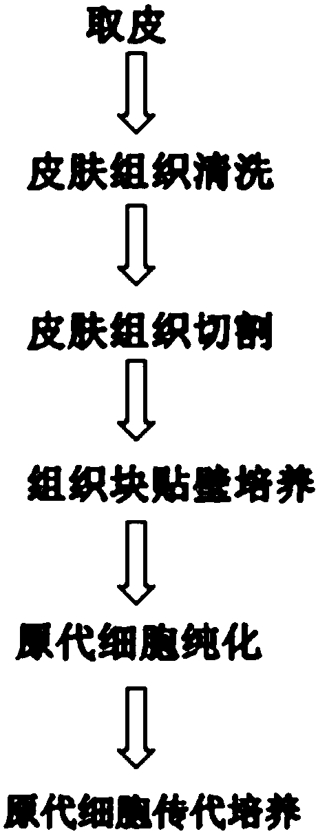
[0042] (1) Isolation of human autologous fibroblasts
[0043] 1) Under a sterile environment, use a sterile skin biopsy puncher to take 3mm skin tissue behind the ear;
[0044] 2) Remove the skin and immediately transfer it to a biological safety cabinet, and rinse it with physiological saline (containing 2% penicillin and streptomycin mixed solution) for 3-5 times;
[0045] 3) Under a stereo microscope, use micro tweezers and a surgical blade to evenly cut 6-9 small pieces of the cleaned skin tissue;
[0046] 4) Evenly spread the cut skin tissue into a 3.5 cm culture dish coated overnight with 1 ml of 0.5% Matrigel (manufacturer: BD; product number: 356234) with an interval of 2 cm between the tissue pieces; add 400 ul of fibroblast culture medium ( Manufacturer: PromoCell GmbH; Item No.: C-23010) placed at 37°C, 5% CO 2 Primary cell culture was carried out in the incubator, and 20...
Embodiment 2
[0049] Embodiment 2: autologous fibroblast identification
[0050] (1) Viability detection of autologous fibroblasts
[0051] Separate the P0 generation autologous fibroblasts in Example 1 to make a single cell suspension, draw 20 μl of the upper cell suspension into a test tube, add 20 μl of 0.4% trypan blue, mix well, and count with a cell counting board after 3 minutes; Observed under the microscope, the dead cells were light blue, and the live cells were rejected, and counted under a 200×high magnification microscope; data analysis, cell survival rate=(total cell number-dead cell number) / total cell number×100%.
[0052] Results: In this experiment, P0 generation autologous fibroblasts were taken for trypan blue exclusion test for three consecutive times, and the viable cell rates of the three checks were 98%, 99%, and 99% respectively.
[0053] (2) Growth curve of autologous fibroblasts
[0054] 1) Take the P5 autologous fibroblasts amplified in Example 1, digest them wi...
Embodiment 3
[0078] Embodiment 3: Determination of collagen content (hydroxyproline) in autologous fibroblast culture fluid
[0079] Since the collagen synthesized by the cultured cells is directly secreted into the culture medium, the content of hydroxyproline (HYP) in the culture medium can indirectly reflect the amount of collagen synthesized by the cultured cells. The culture supernatants of autologous fibroblasts were collected at different time points, and the HYP content was determined with a hydroxyproline test kit.
[0080] For the operation steps, refer to the instructions of Hydroxyproline Detection Kit (manufacturer: Nanjing Jiancheng Bioengineering Research Institute; article number: A030-1):
[0081]
blank tube
standard tube
Assay tube
Double distilled water (ml)
0.25
5ug / ml standard application solution
0.25
Fibroblast culture supernatant (ml)
0.25
Digestive fluid (ml)
0.05
0.05
0.05 ...
PUM
 Abstract
Description
Claims
Application Information
 - R&D
- Intellectual Property
- Life Sciences
- Materials
- Tech Scout
- Unparalleled Data Quality
- Higher Quality Content
- 60% Fewer Hallucinations
Browse by: Latest US Patents, China's latest patents, Technical Efficacy Thesaurus, Application Domain, Technology Topic, Popular Technical Reports.
© 2025 PatSnap. All rights reserved.Legal|Privacy policy|Modern Slavery Act Transparency Statement|Sitemap|About US| Contact US: help@patsnap.com