Self-assembled nucleic-acid aptamer/protein composite nano probe, preparation method, kit and application thereof

A nucleic acid aptamer and protein complex technology, applied in the field of molecular biology, to achieve the effects of low detection limit, improved capture efficiency, and wide linear range

Active Publication Date: 2019-03-15
ZHENGZHOU UNIV
View PDF7 Cites 19 Cited by
  • Summary
  • Abstract
  • Description
  • Claims
  • Application Information

AI Technical Summary

Problems solved by technology

However, rare tumor cell detection strategies based on Aptamer binding to proteins

Method used


Image

  • Self-assembled nucleic-acid aptamer/protein composite nano probe, preparation method, kit and application thereof
  • Self-assembled nucleic-acid aptamer/protein composite nano probe, preparation method, kit and application thereof
  • Self-assembled nucleic-acid aptamer/protein composite nano probe, preparation method, kit and application thereof

Examples


Embodiment 1

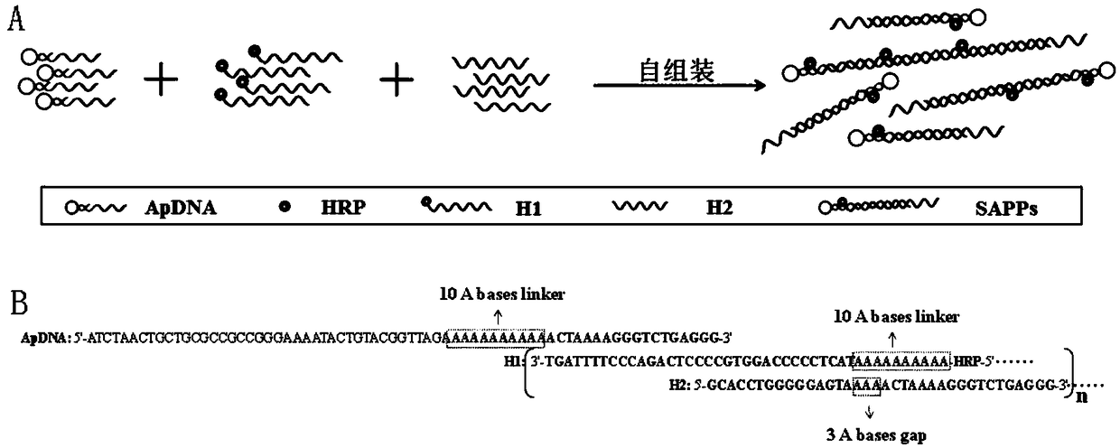
[0057] Preparation of self-assembled nucleic acid aptamer / protein composite nanoprobe: mix 75nmol / L hybridization probe 1, 150nmol / L hybridization probe 2 and 150nmol / L hybridization probe 3 and add to the vortex mixer, vortex at room temperature Spin hybridization for 1 h to obtain the self-assembled nucleic acid aptamer / protein composite nanoprobe. The preparation process is as follows: figure 1 shown.

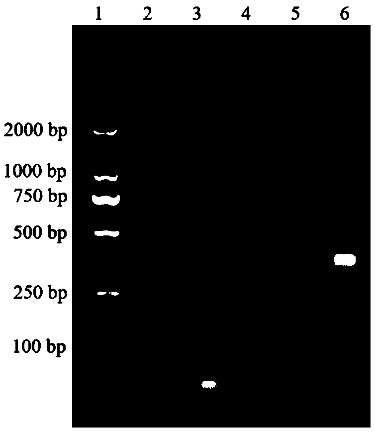
[0058] Characterization of hybridization results: Dilute ApDNA, H1DNA, and H2DNA to a concentration of 2 μmol / L, respectively; then take 10 μL of the above solution, 10 μL of a mixed solution of 1 μmol / L ApDNA, 2 μmol / L H1, and 2 μmol / L H2, respectively, and carry out 2% agar Sugar gel electrophoresis, the electrophoresis conditions are voltage 100V, time 1h, and finally gel imaging, such as figure 2 shown, where figure 2 Middle lane 1: primer; lane 2: binding buffer; lane 3: 2 μmol / L ApDNA; lane 4: 2 μmol / L H1DNA; lane 5: 2 μmol / LH2; 1μmol / L, 2μmol / L and 2μmol / L. Depe...

Embodiment 2

[0060] The preparation method of Sgc8c-MNPs comprises the following steps:

[0061] A: Wash magnetic beads: Take 300 μL 2mg / mL magnetic beads (MNPs) in a 1.5 mL low adsorption centrifuge tube, magnetically separate, and discard the supernatant. Then add 300 μL of PBS buffer solution, mix thoroughly, magnetically separate, discard the supernatant, and wash 3 times in total.

[0062] B: Combination of MNPs and Sgc8c: Add 500 μL of 400 nmol / L Sgc8c (another portion of Sgc8c with the same concentration, marked as pre) to the above-mentioned low-adsorption centrifuge tube, and mix well. Vortex reaction at room temperature for 30 minutes, magnetically separate, save the supernatant, marked as post, wash the magnetic beads twice with PBS, 500 μL each time, save the supernatant separately, marked as wash 1 and wash 2;

[0063] C: The MNPs modified with Sgc8c (Sgc8c-MNPs) were resuspended in 600 μL of immunomagnetic bead preservation solution, and stored at 4°C for use.

[0064] Char...

Embodiment 3

[0071] Capture of target cells by Sgc8c-MNPs in a single CCRF-CEM suspension:

[0072] (1) Wash cells: wash CCRF-CEM 3 times with binding buffer, then resuspend for 10 6 cells / mL cell suspension;

[0073] (2) Cell staining: 5 μL of 2 mmol / L DiD dye was added to each ml of CCRF-CEM cell suspension, and incubated at 37° C. for 5 min. Wash cells 3 times with binding buffer;

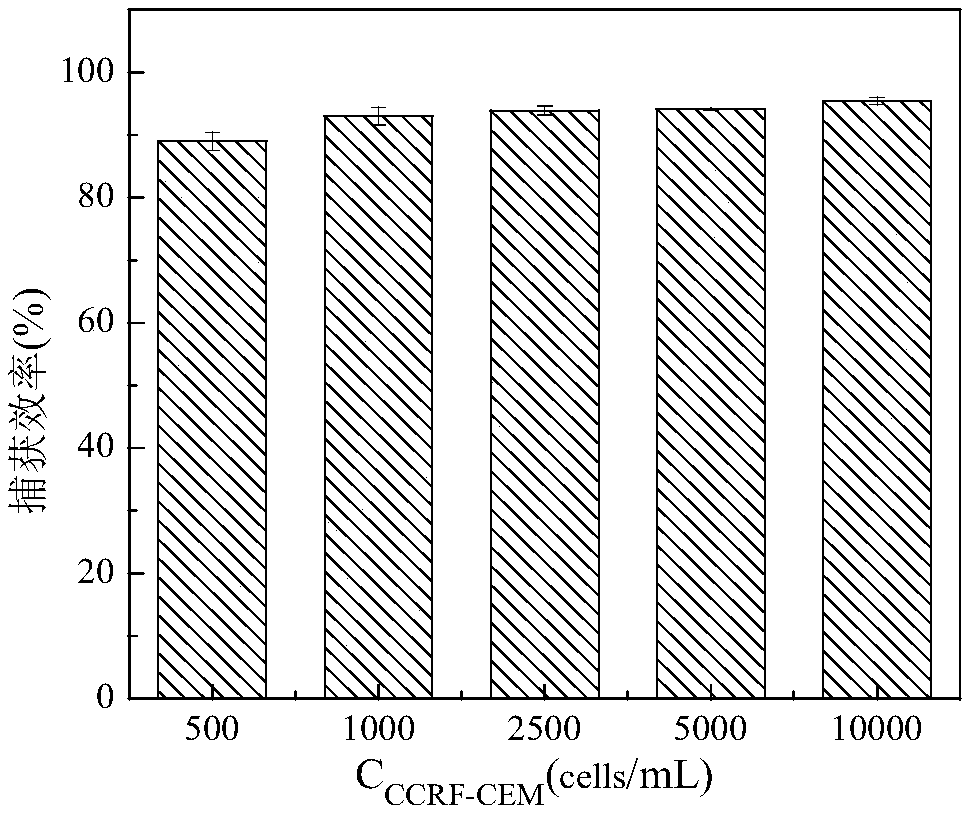
[0074] (3) Sgc8c-MNPs capture CCRF-CEM: Dilute the stained cells into five different concentrations: 500, 1000, 2500, 5000, 10000 cells / mL. Take 1 mL of different concentrations of CCRF-CEM and mix them with Sgc8c-MNPs (the amount of Sgc8c-MNPs is 20 μL of stock solution), incubate at 4°C for 20 min, magnetically separate, and collect the supernatant. Centrifuge at 1000r / min for 5min, and disperse the cell pellet in 100μL binding buffer;

[0075] (4) Cell counting: Under the laser confocal microscope, the CCRF-CEM in the supernatant was counted for fluorescence, and the capture efficiency (Capture effici...


PUM

PropertyMeasurementUnit
particle sizeaaaaaaaaaa

Abstract

The invention discloses a self-assembled nucleic-acid aptamer/protein composite nano probe, a preparation method, a kit and application thereof. According to the invention, three DNA probes, one of which is a DNA probe containing a nucleic-acid aptamer sequence and another is marked with horseradish peroxidase, are hybridized to prepare a self-assembled nucleic-acid aptamer/protein composite nanoprobe; and magnetic nano-particles functionalized by a nucleic-acid aptamer are used as magnetic separation solid-phase carriers for specific capture of human acute lymphoblastic leukemia T lymphocytes (CCRF-CEM) in blood, to improve the capture efficiency of target substances. When the magnetic nano-particles are used in combination with the self-assembled nucleic-acid aptamer/protein composite nano probe, the detection of CCRF-CEM has a wide linear range, a low detection limit, and high precision and accuracy, and high-sensitivity, high-specificity and rapid detection of CCRF-CEM in peripheral blood samples can be realized, which has great significance for auxiliary diagnosis, curative effect evaluation and prognosis judgment of leukemia.

Description

technical field [0001] The invention relates to the technical field of molecular biology, in particular to a self-assembled nucleic acid aptamer / protein composite nanoprobe, a preparation method, a kit and an application thereof. Background technique [0002] Currently, self-assembly, as a powerful technique to spontaneously organize or aggregate functional building blocks into multifunctional materials in a controllable manner, has been successfully used in sensing and drug loading in biomedicine. In particular, self-assembly based on nucleic acid hybridization regulation, due to the diversity of nucleic acid structure and function, and the advantages of programmable design of its sequence, makes it useful in the orderly construction of nanomaterials, nucleic acid, enzyme activity, heavy metal ion detection, etc. It has broad application prospects. In recent years, the construction and application of nucleic acid assemblies using nucleic acid aptamers (Aptamers) as affinit...

Claims


Application Information

Patent Timeline
Patent Type & Authority Applications(China)
IPC IPC(8): G01N21/76
CPCG01N21/76
Inventor 何磊良邓猛聪赵梦瑶沙吉旦·布拉了高宁振刘焕岳帅汪俊朱星晨
Owner ZHENGZHOU UNIV
Why Eureka
Try Eureka