Construction method and application of in-vitro respiratory exposure model

A construction method, external respiration technology, applied in artificial cell constructs, biochemical equipment and methods, epidermal cells/skin cells, etc. The method that can solve cytotoxicity cannot simulate the lungs well, and cannot simulate the lungs well To prevent lung collapse and maintain the balance of surface tension

Active Publication Date: 2019-03-19
QINGDAO UNIV
View PDF3 Cites 4 Cited by
  • Summary
  • Abstract
  • Description
  • Claims
  • Application Information

AI Technical Summary

Problems solved by technology

But this exposure model is not a good simulation of the real environment of the lungs
The method of directly adding toxic substances or drugs to the medium to evaluate cytotoxicity cannot simulate the real environment of the lungs well, ignoring the influence of the real lung microenvironment on toxic substances or drugs

Method used


Image

  • Construction method and application of in-vitro respiratory exposure model
  • Construction method and application of in-vitro respiratory exposure model
  • Construction method and application of in-vitro respiratory exposure model

Examples


Embodiment 1

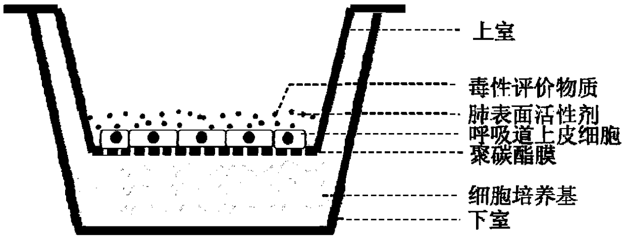
[0030] Human lung epithelial cells Beas-2B were cultured in vitro. After the cells grew to the logarithmic growth phase, the medium was removed, washed three times with phosphate buffer, and then digested with trypsin for 3 minutes. After digestion, add MEM medium containing fetal bovine serum to stop, add fresh medium after centrifugation and resuspend, according to 5×10 4 / cm 2 The cell density was inoculated onto the perforated membrane material in the upper chamber of the 3470 model co-cultivation system, and placed in the cell incubator to continue culturing for 24 hours;

[0031] Add 500 microliters of MEM cell culture medium containing fetal bovine serum to the lower chamber of the co-culture model, nest the upper chamber inoculated with the cells into the lower chamber, carefully aspirate the liquid in the upper chamber of the co-culture system, and then place it in the upper chamber Add 100 microliters of bovine lung surfactant and continue culturing for 12 hours;

...

Embodiment 2

[0034] Human lung epithelial cells 16HBE were cultured in vitro. After the cells grew to the logarithmic growth phase, the medium was removed, washed three times with phosphate buffer, and then digested with trypsin for 5 minutes. After digestion, add MEM medium containing fetal bovine serum to terminate, add fresh medium after centrifugation, and resuspend according to 1×10 5 The cell density was inoculated onto the perforated membrane material in the upper chamber of the 3491 model co-cultivation system, and placed in the cell incubator to continue culturing for 24 hours;

[0035]Add 500 microliters of MEM cell culture medium containing fetal calf serum to the lower chamber of the co-culture model, and nest the upper chamber where the cells were inoculated into the lower chamber; carefully aspirate the liquid in the upper chamber of the co-culture system, and then place it in the upper chamber Add 100 microliters of bovine lung surfactant and continue culturing for 12 hours;...

Embodiment 3

[0039] Human lung epithelial cells 16HBE were cultured in vitro. After the cells grew to the logarithmic growth phase, the medium was removed, washed three times with phosphate buffer, and then digested with trypsin for 5 minutes. After digestion, add MEM medium containing fetal bovine serum to terminate, add fresh medium after centrifugation, and resuspend according to 1×10 5 The cell density was inoculated onto the perforated membrane material in the upper chamber of the 3491 model co-cultivation system, and placed in the cell incubator to continue culturing for 24 hours;

[0040] Add 500 microliters of MEM cell culture medium containing fetal calf serum to the lower chamber of the co-culture model, and nest the upper chamber where the cells were inoculated into the lower chamber; carefully aspirate the liquid in the upper chamber of the co-culture system, and then place it in the upper chamber Add 100 microliters of bovine lung surfactant and continue culturing for 12 hours...


PUM

PropertyMeasurementUnit
pore sizeaaaaaaaaaa

Abstract

The invention provides a construction method of an in-vitro respiratory exposure model, comprising the steps of inoculating respiratory epithelial cells to a porous membrane material on a co-culture system, culturing until the cells attach to a wall, wherein the membrane material divides the co-culture system into an upper chamber and a lower chamber; adding a cell culture medium into the lower chamber of the co-culture system, adding a lung surfactant to the upper chamber, and continuing to culture for 0.5-2 hours; adding a toxic material or a drug into the upper chamber to carry out exposure, and evaluating toxicological indexes of the respiratory epithelial cells. Application of the construction method of the in-vitro respiratory exposure model in environmental pollutant or drug evaluation is also provided.

Description

technical field [0001] The invention belongs to the technical field of biomedical engineering, and in particular relates to a construction method and application of an in vitro respiratory exposure model. Background technique [0002] The respiratory system is an organ system for the human body to exchange gas with the external environment, including the gas passage nose, throat, trachea, bronchi, and the gas exchange organ lung. Due to the special structure and function of the respiratory system, some gaseous pollutants or particles can enter the human body through the respiratory route such as the nose or mouth, thereby endangering human health. In addition, some lung diseases, such as asthma, chronic obstructive pulmonary disease, etc., can also be administered through the respiratory tract during treatment. After pollutants or drugs enter the respiratory tract, they will have a certain impact on the epithelial cells of the respiratory tract, thereby further affecting th...

Claims


Application Information

Patent Timeline
Patent Type & Authority Applications(China)
IPC IPC(8): C12N5/071C12Q1/02
CPCC12N5/0625G01N33/5044
Inventor 唐敬龙王红梅程文婷尹慧敏
Owner QINGDAO UNIV
Why Eureka
Try Eureka