Gynecological tumor cell combined quantitative detection card, preparation method thereof, and using method thereof

A quantitative detection technology for gynecological tumors, applied in measuring devices, scientific instruments, biological tests, etc., can solve the problems of inability to accurately determine whether there is metastasis, inability to achieve real-time dynamic monitoring, inability to reflect heterogeneity, etc., to achieve detection time Short, easy to popularize and use, and increase the effect of response sensitivity

Inactive Publication Date: 2019-05-14
周辉
View PDF0 Cites 1 Cited by
  • Summary
  • Abstract
  • Description
  • Claims
  • Application Information

AI Technical Summary

Problems solved by technology

For example, by using serum gynecological malignant tumor cell identification, due to individual differences, some gynecological malignant tumors lack highly specific markers and cannot be effectively detected, and their reliability is poor; However, even with the most advanced and accurate imaging detection methods, the detection diameter of gynecological malignant tumors must be at least 2-3 mm, that is, gynecological malignant tumors smaller than this diameter range are often not found by imaging examinations, so It can only be applied to the detection of advanced cancer; histopathological sections are often identified based on puncture or gynecological malignant tumor tissue taken out after surgery, and the technical requirements are high, and it cannot be obtained repeatedly, for a long time or at any time. In addition, the The method is also invasive to a certain extent and cannot accurately determine whether there is metastasis; endoscopic exploration is relatively invasive, and the patient's willingness to cooperate is low. Single-site sampling cannot reflect heterogeneity, and real-time dynamic monitoring cannot be achieved. , can only simply confirm the existence of gynecological malignant tumors, but it is impossible to determine whether the cells of gynecological malignant tumors have infiltrated and metastasized

Method used


Image

  • Gynecological tumor cell combined quantitative detection card, preparation method thereof, and using method thereof
  • Gynecological tumor cell combined quantitative detection card, preparation method thereof, and using method thereof

Examples


Embodiment 1

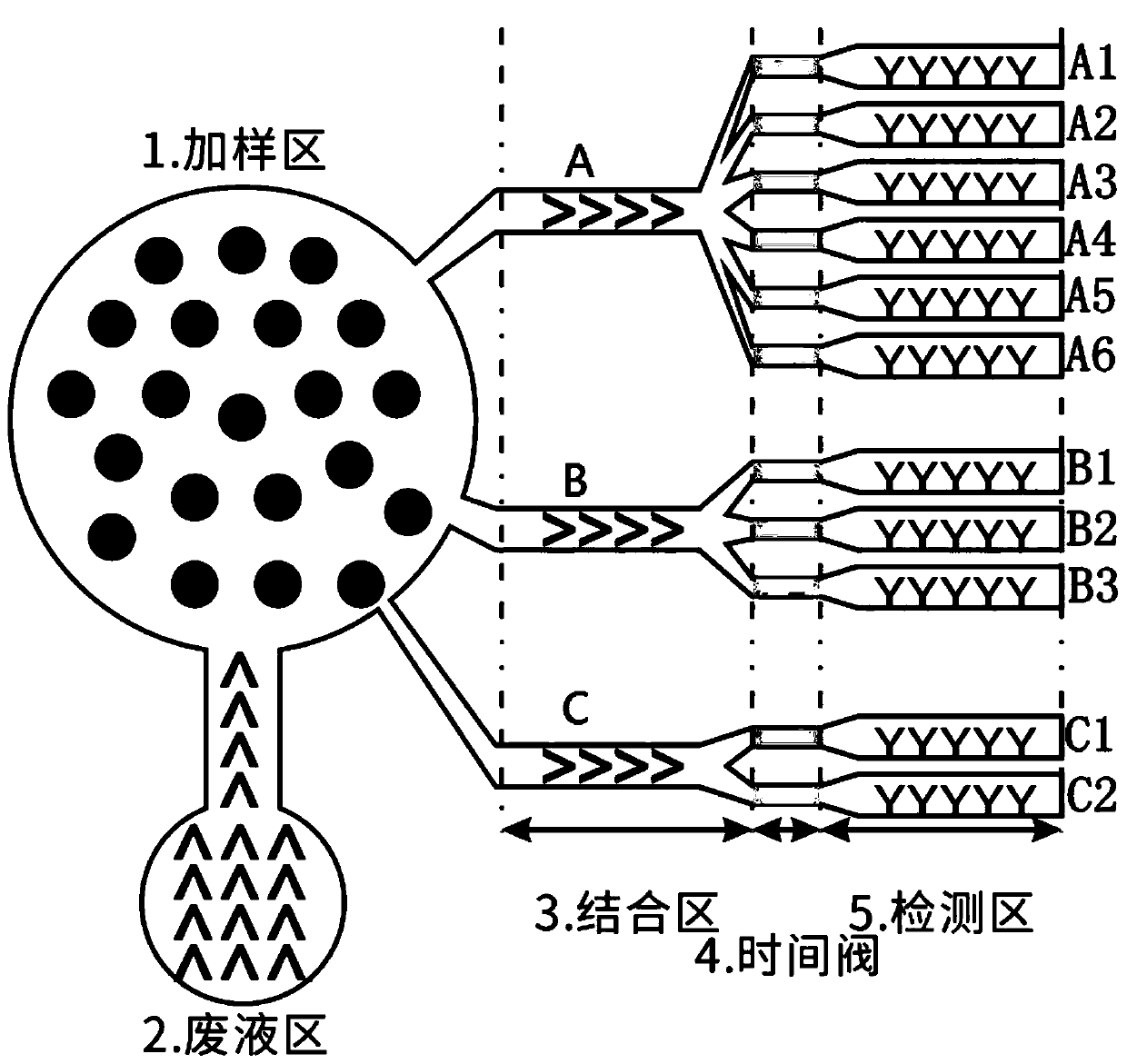
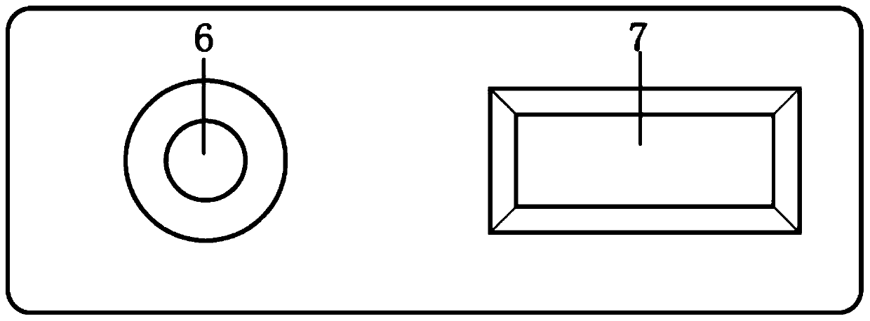
[0071] A combined quantitative detection card for gynecological tumor cells, comprising a bottom card and a face card, the bottom card is provided with: a sample loading area, a binding area, a microchannel, a time valve, a detection area and a waste liquid area, the sample loading area Located at one end of the microchannel, the binding area is connected to a plurality of detection areas through the microchannel and a time valve and located at the other end of the microchannel, and the waste liquid area is connected to the sample loading area through the microchannel;

[0072] There are multiple binding regions.

[0073] The binding area A is connected to the detection areas A1, A2, A3, A4, A5, and A6 through microchannels and time valves;

[0074] The binding area B is connected to the detection areas B1, B2, and B3 through microchannels and time valves;

[0075] The binding area C is connected to the detection areas C1 and C2 through a microchannel and a time valve.

[00...

Embodiment 2

[0101] A method for preparing a combined quantitative detection card for gynecological tumor cells, comprising the following steps:

[0102] (1) Preparation of bottom card:

[0103] (1-1) Drawing: draw the mask layout through the drawing software according to the design of the microchannel pattern;

[0104] (1-2) Mask: After the mask layout is made, print it on a transparent film to make a photolithography mask plate, and use this mask plate to make a silicon wafer mold through a photolithography process;

[0105] (1-3) Photolithography: plate a layer of barrier layer on the surface of the substrate, and then evenly throw a layer of photosensitive material-photoresist on the barrier layer with a glue machine; prepare the required film on the photomask Channel pattern; cover the photomask on the substrate, irradiate the substrate coated with photoresist with ultraviolet light, and the photoresist undergoes photochemical reaction; remove the exposed photoresist by chemical meth...

Embodiment 3

[0113] The method for using the combined quantitative detection card for gynecological tumor cells comprises the following steps:

[0114] (a) The aqueous solution A containing dextran, trehalose and gelatin was added to the aqueous solution B containing polyethylene glycol to form a fibril network, and then 1 wt% egg white lysozyme was added for heat incubation at 60°C, the fibrils The network is transformed into mature fibrils, and finally the serum standard is added to it, and the mature fibrils are used to wrap the serum standard, and water-in-water micro-droplets with a diameter of 8 μm can be prepared and prepared into a series of concentrations of more than 5 , use several detection cards of the same batch to detect the standard solution of each concentration, take the fluorescence intensity of the detection area as the ordinate, and the concentration of the serum standard solution as the abscissa, draw a standard curve and save the standard curve in the multi-color fluo...


PUM

PropertyMeasurementUnit
pore sizeaaaaaaaaaa
diameteraaaaaaaaaa
diameteraaaaaaaaaa

Abstract

The invention discloses a gynecological tumor cell combined quantitative detection card, a preparation method thereof, and a using method thereof. The gynecological tumor cell combined quantitative detection card comprises a bottom card and a surface card; the bottom card is provided with a sample adding zone, bonding zones, microchannels, time valves, detection zones, and waste liquid zones, wherein the sample adding zone is positioned at one end of the microchannels, the bonding zones are connected with the multiple detection zones through the microchannels and the time valves and are positioned at the other end of the microchannels, the waste liquid zones are connected to the sample adding zone through the microchannels, and the bottoms of the bonding zones and the detection zones are both parallelly connected with the microchannels; and the microchannels are connected with the time valves. According to the gynecological tumor cell combined quantitative detection card, the preparation method thereof, and the using method thereof,the flow rate and the flow quantity of a to-be-tested sample are controlled by the microfluidic technology, gynecological tumor cells with the same marker enter the detection zones through a microchannel, so that mutual interference is avoided, thus the error is reduced, and reaction sensitivity is increased; and the to-be-tested sample is prepared into water-in-water micro-droplets, so that the detection limit is increased.

Description

technical field [0001] The invention relates to the technical field of combining microfluidic technology and immunofluorescence microsphere immunochromatography technology, in particular to a combined quantitative detection card for gynecological tumor cells and its preparation method and use method. Background technique [0002] Over the years, gynecological malignancies have been the main diseases that threaten the health and life of women. Ovarian cancer, cervical cancer, and endometrial cancer are the three most common gynecological malignancies. In recent years, the incidence has been increasing year by year and younger. The current methods for clinical examination of gynecological malignant tumors, such as serum gynecological malignant tumor cell identification, medical image scanning, histopathological section, endoscopic exploration, etc. For example, by using serum gynecological malignant tumor cell identification, due to individual differences, some gynecological m...

Claims


Application Information

Patent Timeline
Patent Type & Authority Applications(China)
IPC IPC(8): G01N33/558G01N33/58G01N33/533G01N33/574
Inventor 周辉
Owner 周辉
Why Eureka
Try Eureka