Preparation method of 3D tissue engineering cell ring

A tissue engineering and cell technology, applied in the field of cell engineering, can solve the problems of geometric size gap, complicated operation process, scaffolds affecting cell growth and differentiation, etc.

Pending Publication Date: 2019-12-13
吴宏伟 +1
View PDF4 Cites 6 Cited by
  • Summary
  • Abstract
  • Description
  • Claims
  • Application Information

AI Technical Summary

Problems solved by technology

Two-dimensional cell culture models are still far from accurately reproducing the function of cells in tissues
The cell scaffold is mainly to construct a porous structure in three-dimensional space for cell attachment and growth. Cells attach to the scaffold for three-dimensional growth and migration. The main scaffold materials include collagen and hydrogel. The three-dimensional culture technology that does not require scaffolds is mainly through Physical methods suspend adherent cells in the medium to achieve the purpose of three-dimensional culture. At present, the main technologies include microcarrier, magnetic levitation, hanging drop plate and magnetic three-dimensional bioprinting. These methods are relatively complicated to operate and require a large initial investment. At present, the vast majority of three-dimensional cultures rely on scaffold materials
The current hanging drop culture method can only grow microspheres with a particl

Method used


Image

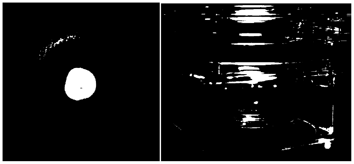
  • Preparation method of 3D tissue engineering cell ring
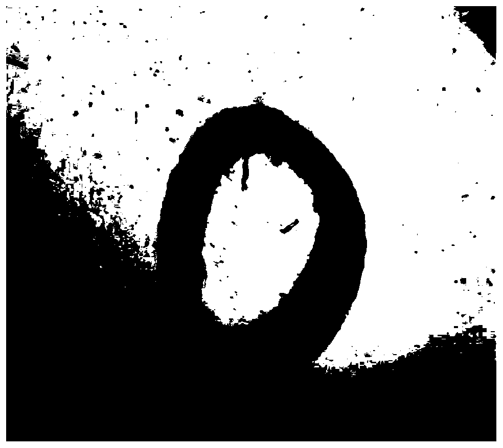
  • Preparation method of 3D tissue engineering cell ring
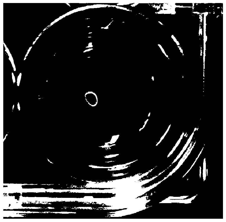
  • Preparation method of 3D tissue engineering cell ring

Examples


Embodiment 1

[0117] Example 1: Human osteosarcoma 143b cell ring (the preparation method of human lung cancer A549 cell ring and human nasopharyngeal carcinoma S18 cell ring is the same as that of osteosarcoma)

[0118] A preparation method of human osteosarcoma 143b cell ring, comprising the following steps:

[0119] a. Prepare ring culture medium: add 0.25% methylcellulose to high-sugar DMEM basic medium, add 10% FBS, 10ng / mlbFGF, 10ng / ml PDGF, 20ng / ml TGFβ, 10uM Y-27632;

[0120] b. Put the low-temperature sterilized cell ring culture mold into a 24-well culture plate;

[0121] c. Add 40 microliters of anti-adhesion coating to the culture well and incubate at room temperature for 30 minutes;

[0122] d. Aspirate the anti-adhesion coating, and rinse the groove of the culture well with room temperature DMEM medium for 30 minutes;

[0123] e. Suck out the DMEM base, and the mold is ready for use;

[0124] f. After the 143b osteosarcoma and human epidermal fibroblasts grow to 95% saturatio...

Embodiment 2

[0132] Example 2: Human giant cell tumor of bone stromal cell ring (the preparation method of rat adipose stem cell ring is the same as that of giant cell tumor of bone)

[0133] A method for preparing a human giant cell tumor stromal cell ring, comprising the following steps:

[0134] a. Prepare ring culture medium: add 0.25% methylcellulose to high-sugar DMEM basic medium, add 10% FBS, 10ng / mlbFGF, 10ng / ml PDGF, 20ng / ml TGFβ, 10uM Y-27632;

[0135] b. Put the low-temperature sterilized cell ring culture mold into a 24-well culture plate;

[0136] c. Add 40 microliters of anti-adhesion coating to the culture well and incubate at room temperature for 30 minutes;

[0137] d. Aspirate the anti-adhesion coating, and rinse the groove of the culture well with room temperature DMEM medium for 30 minutes;

[0138] e. Suck out the DMEM base, and the mold is ready for use;

[0139] f. After the 143b giant cell tumor cells grow to 95% saturation, digest the cells with 0.5% trypsin to...


PUM


Abstract

The invention discloses a preparation method of a 3D tissue engineering cell ring. By utilizing a relatively narrow growth space and a surface non-adhesion characteristic of a culture mold, added cells are self-assembled into a ring in a culture solution for promoting cell aggregation and leave the surface of a cell culture medium, and a macroscopic tissue block or organoid with a three-dimensional structure is formed. A naturally-formed extracellular matrix exists between the cells prepared by the method, the exogenous scaffold interference does not exist, and the communication and the information exchange between the cells are more complete and direct so as to reduce the difference between a culture environment and an in-vivo environment and more truly simulate an in-vivo cell growth environment. Moreover, the prepared cell ring is a cell body with a regular annular structure, can simulate the in-vivo cell microenvironment, links a 2D cell culture system with the research of tissuesand organs, and is beneficial to research of organoids.

Description

technical field [0001] The invention relates to the field of cell engineering, in particular to a method for preparing 3D tissue engineering cell rings. Background technique [0002] Traditional medical research and cell biology research mostly adopt the two-dimensional cell culture model, which provides a flat culture plane. However, under physiological conditions, tissue cells grow in three-dimensional space, and cells not only contact each other, but also contact with supporting structures such as extracellular matrix. The two-dimensional cell culture model cannot really reflect the extracellular matrix in the tissue, so many physiological reactions in this model, such as receptor expression, transcription expression, cell migration and apoptosis, have been confirmed to be consistent with those that occur in actual organs or tissues. The situation is different. Moreover, the entire process of cell division, proliferation, migration and apoptosis is precisely regulated, ...

Claims


Application Information

Patent Timeline
IPC IPC(8): C12N5/071C12N5/09C12M3/00C12M1/00
CPCC12N5/0602C12N5/0693C12M23/02C12N2513/00
Inventor 吴宏伟黄钢许彦钟午李先安
Owner 吴宏伟
Why Eureka
Try Eureka