Drug-loading nanofiber membrane for preventing nasal infection and adhesion and preparation method thereof

A drug-loaded nano-fiber membrane technology, applied in fiber treatment, fiber chemical characteristics, rayon manufacturing, etc., can solve the problems of limited drug-loading capacity and unsatisfactory adhesion effect, so as to avoid adverse reactions, prevent repeated infections, good preventive effect

Inactive Publication Date: 2019-12-27
SHANGHAI CHANGHAI HOSPITAL
View PDF9 Cites 2 Cited by
  • Summary
  • Abstract
  • Description
  • Claims
  • Application Information

AI Technical Summary

Problems solved by technology

[0004] The existing auxiliary artificial materials can be divided into non-absorbable materials and absorbable materials. The disadvantage of using non-absorbable materials is that a second operation is required; while using absorbable materials such as collagen and animal derivatives is limited by the material Due to the limitation of selection and drug loading capacity, the effect of wound healing, maintaining patency, preventing infection and adhesion is not ideal

Method used


Image

  • Drug-loading nanofiber membrane for preventing nasal infection and adhesion and preparation method thereof
  • Drug-loading nanofiber membrane for preventing nasal infection and adhesion and preparation method thereof
  • Drug-loading nanofiber membrane for preventing nasal infection and adhesion and preparation method thereof

Examples


Embodiment 1

[0055] Preparation of PLLA-PVP membrane loaded with clarithromycin in embodiment 1

[0056] Add 1g PLLA, 0.01g PVP and 0.1g clarithromycin to the mixture of 4g dichloromethane and 2g N,N-dimethylformamide, stir constantly for 1 hour to completely dissolve PLLA and other materials; The spinning solution was sonicated for 10 min in a water bath to eliminate any air bubbles. The mixed solution after sonication was placed in a 10 mL syringe, the diameter of the syringe needle was 0.8 mm, the voltage was fixed at 15 kV, the distance between the needle and the collector was set at 15 cm, and the solution feeding rate was 0.2 mL / Electrospinning was carried out under the condition of 10 minutes, and an electrospun fiber membrane loaded with clarithromycin (CLA-PLLA-PLLA, clarithromycin concentration 10%) was produced.

[0057] And, the PLLA film was produced according to the similar production steps as above; except that only 1 g of PLLA was dissolved in an organic solvent when prep...

Embodiment 2

[0058] Embodiment 2 physical performance detection

[0059] (1) Scanning electron microscope observation

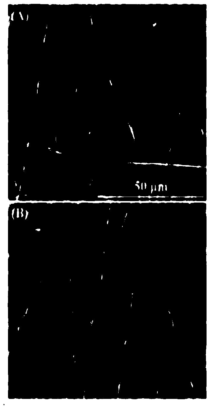
[0060] With prepared PLLA film and CLA-PLLA-PVP film (kind see figure 2 ) was collected on the surface of aluminum foil, followed by vacuum drying for 24 hours, and the morphology of PLLA and CLA-PLLA-PVP fiber membrane was observed by scanning electron microscope.

[0061] image 3Scanning electron micrographs of the PLLA film and the CLA-PLLA-PVP film prepared in Example 1, A is the PLLA film, and B is the CLA-PLLA-PVP film. According to the results of scanning electron microscopy, there is no obvious difference in the fiber morphology of PLLA membrane and CLA-PLLA-PVP membrane. Both of them have smooth surface, similar fiber diameter and porosity. 30% to 95%. It can be seen that the drug loading process of the present invention basically has no effect on the appearance of these fibers.

[0062] (2) Water contact angle measurement

[0063] Water contact angle (wa...

Embodiment 3

[0069] Embodiment 3 in vitro drug experiment

[0070] (1) In vitro drug release ability

[0071] Each 200 mg of CLA-PLLA-PVP fiber membrane was immersed in 50 mL of phosphate-buffered saline (PBS, pH 7.4) solution, placed in a thermostat shaking water bath at a shaking speed of 50 cycles / min. On the 15th, 30th, 45th, and 60th days, 1.0 mL of sustained-release solution medium was taken out for detection, and 1.0 mL of fresh PBS solution was added at the same time. The PBS solution taken out was analyzed by high-performance liquid chromatography (HPLC), and the stationary phase was analyzed at 25°C using an ODS column; the mobile phase was a potassium dihydrogen phosphate solution / methanol (400:600v / v) system; the flow rate was 1.0mL / min, the detection wavelength is 210nm.

[0072] For drug release see Figure 6 , from the drug release curve of the CLA-PLLA-PVP film, it can be seen that clarithromycin can be released slowly and steadily for two months, and a clarithromycin r...


PUM

PropertyMeasurementUnit
diameteraaaaaaaaaa
tensile strengthaaaaaaaaaa
thicknessaaaaaaaaaa

Abstract

The invention provides a drug-loading nanofiber membrane for preventing nasal infection and adhesion and a preparation method thereof; the drug-loading nanofiber membrane comprises poly-L-lactic acid,polyvinylpyrrolidone and clarithromycin; the drug-loading nanofiber membrane has a fiber diameter less than 10 micrometers. According to the invention, after being loaded with clarithromycin, the poly-L-lactic acid and PVP composite fiber membrane can be implanted and adhered to the nasal cavity and paranasal sinuses to continuously release the clarithromycin, can degrade by itself and can play antibacterial, anti-inflammatory and anti-adhesion roles after chronic rhinosinusitis and nasosinusitis surgeries, thereby preventing postoperative repeated infection, adhesion and other complications.The drug-loading nanofiber membrane provided by the invention can be used for preventing infection and adhesion of nasal cavities and sinuses, and has the advantages of convenience in use, avoidanceof repeated drug administration, avoidance of adverse reactions of systemic drug administration and the like.

Description

technical field [0001] The invention relates to the technical field of medical materials, in particular to a drug-loaded nanofiber membrane for preventing nasal infection adhesion and a preparation method thereof. Background technique [0002] Chronic rhinosinusitis is a common clinical disease. The clinical definition refers to chronic inflammation of the nasal cavity and sinus mucosa. The nasal symptoms last for more than 12 weeks, and the symptoms are not completely relieved or even aggravated. Nasal endoscopic surgery is an important treatment, and the key to nasal endoscopic surgery is to restore the mucociliary system and reconstruct the airway. However, fresh wounds in the local mucosa will inevitably occur during the operation. If some factors such as re-infection and local sinus stubborn inflammation are affected during the recovery process, complications such as repeated infections and adhesions may occur in the later recovery period, which can be serious. Affect ...

Claims


Application Information

Patent Timeline
Patent Type & Authority Applications(China)
IPC IPC(8): D04H1/728D01D5/00D01F1/10D01F6/92
CPCD01D5/0007D01D5/003D01D5/0061D01F1/10D01F1/103D01F6/92D04H1/728D10B2401/13D10B2509/00
Inventor 唐海红郑宏良崔文国张涛陈争明唐梓荧
Owner SHANGHAI CHANGHAI HOSPITAL
Why Eureka
Try Eureka