Interface targeting type mitochondrial probe as well as preparation method and application thereof

A mitochondrial, targeting technology, applied in chemical instruments and methods, instruments, analytical materials, etc., can solve problems such as inability to live cells, mitochondrial cell damage, etc., and achieve the effects of low cytotoxicity, intuitive biological detection reagents, and good application prospects.

Inactive Publication Date: 2020-07-03
JIANGXI SCI & TECH NORMAL UNIV
View PDF3 Cites 9 Cited by
  • Summary
  • Abstract
  • Description
  • Claims
  • Application Information

AI Technical Summary

Problems solved by technology

However, this type of probe must react to mitochondrial proteins, which will c

Method used


Image

  • Interface targeting type mitochondrial probe as well as preparation method and application thereof
  • Interface targeting type mitochondrial probe as well as preparation method and application thereof
  • Interface targeting type mitochondrial probe as well as preparation method and application thereof

Examples


Embodiment 1

[0039] A method for preparing an interface-targeted mitochondrial probe, comprising:

[0040] (1) Synthesis of 7-diethylamino-coumarin: 4mmol 4-(diethylamino) salicylaldehyde (0.77g) and 10mmol diethyl malonate (1.6g) were dissolved in 30mL absolute ethanol, Add 1 mL of piperidine. The above mixture was stirred at 80 °C for 6 h. After removal of ethanol, 20 mL of concentrated hydrochloric acid and 20 mL of glacial acetic acid were added. The above solution was stirred and refluxed at 120° C. for 10 h. Then the mixture was cooled to room temperature and poured into 50 mL of ice water. Subsequently, the pH of the solution was adjusted to 5.0 with 1.0 mol / L NaOH solution. The mixture was filtered to give a solid which was then dissolved in a small amount of dichloromethane. A large amount of n-hexane was added to obtain the product with a yield of 90%.

[0041] (2) Synthesis of 7-diethylamino-coumarin-3-carbaldehyde: the three-necked flask was placed in an ice bath at 0° C....

Embodiment 2

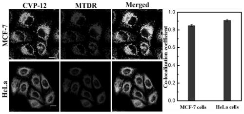
[0045] Example 2 cell (HeLa and MCF-7) culture and staining

[0046] All cell lines were maintained at 37°C in 5% CO 2 cultured in a saturated humidity incubator. Cell lines were cultured in H-DMEM medium containing 10% fetal bovine serum (FBS) and 1% penicillin and streptomycin. CVP-12 was dissolved in DMSO solution to prepare a stock solution with a concentration of 0.1 mM. For live cell staining experiments, cultured cells grown on glass coverslips were stained with 1 μM CVP-12 in culture medium for 10 minutes at 37°C and imaged with a fluorescent microscope.

Embodiment 3

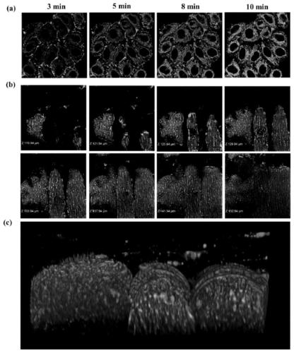
[0047] Example 3 Membrane permeability experiment of CVP-12 staining HeLa cells and muscle tissue

[0048] For foreign organic probes, their self-delivery ability is usually weak due to the strict selectivity of the membrane structure for substances entering the cell. The membrane permeability of the probe is an important parameter for imaging cells and tissues, and the present invention tests the permeability of CVP-12. Such as figure 1 As shown in a, CVP-12 can image mitochondria in 3 minutes, indicating that the probe can enter active HeLa cells in a short time. Then, the present invention further tested the permeability of CVP-12 in tissue by probe imaging depth. After 30min, the image was obtained by optical sectioning of the tissue ( figure 1 b), and obtain the computer-generated 3D reconstruction image ( figure 1 c). The fluorescent signal of mitochondria can still be clearly detected at a depth of 52 μm, and there is no background noise. The above results show th...


PUM


Abstract

The invention discloses an interface targeting type mitochondrial probe as well as a preparation method and application thereof in living cells and tissues, and belongs to the technical field of mitochondrial fluorescent probes. The structural formula of the interface targeting type mitochondrial probe is shown in the specification. The chemical name of the interface targeting type mitochondrial probe is (E)-4-(2-(7-diethylamino-coumarin) vinyl)-1-dodecyl pyridine iodate. The probe is named as CVP-12. The interface targeting type mitochondrial probe has the characteristics of no need of washing, high selectivity, good biocompatibility, excellent membrane permeability and low cytotoxicity, and has good application prospects in the field of biomarkers.

Description

technical field [0001] The invention relates to the technical field of mitochondrial fluorescent probes, in particular to an interface-targeted mitochondrial probe, its preparation method and its application in living cells and tissues. Background technique [0002] As a semi-autonomous organelle in cells, mitochondria can not only provide energy for cells, but also participate in processes such as cell differentiation, cell information transmission, and cell apoptosis, and can regulate cell growth and cell cycle. Mitochondrial dysfunction is therefore responsible for many diseases, such as Parkinson's disease and Alzheimer's disease. Generally, dysfunctional mitochondria are cleared by autophagy to maintain cell health. Therefore, detecting mitochondrial dynamics and mitophagy process is of great help to our understanding of microphysiological changes and mitochondria-related diseases. [0003] Initially, mitochondria were visualized using electron microscopy and radioact...

Claims


Application Information

Patent Timeline
IPC IPC(8): C07D405/06C09K11/06G01N21/64
CPCC07D405/06C09K11/06C09K2211/1029C09K2211/1088G01N21/6428G01N2021/6439
Inventor 徐景坤张革刘芳李斐张新新
Owner JIANGXI SCI & TECH NORMAL UNIV
Why Eureka
Try Eureka