Method for inducing differentiation of adipose derived stem cells into chondrocyte

An adipose stem cell and induction differentiation technology, which is applied in the field of adipose stem cells to induce differentiation into chondrocytes, can solve the problems of reduced expression of chondrocytes marker genes, inability to apply clinical treatment on a large scale, and low efficiency of osteoblast differentiation. Cell adhesion, beneficial to cell proliferation and differentiation, and the effect of improving the efficiency of induction of differentiation

Active Publication Date: 2020-07-28
SHANDONG XINRUI BIOTECH CO LTD
View PDF2 Cites 3 Cited by
  • Summary
  • Abstract
  • Description
  • Claims
  • Application Information

AI Technical Summary

Problems solved by technology

[0006] The microsphere method and the microdrop method are used to culture adipose-derived stem cells to induce differentiation into cartilage. Although it is simple and convenient, and does not require specific biological materials, the cells aggregate and grow during the culture process, which is prone to defects of nutritional deficiencies, resulting in slow cell proliferation and osteogenesis. The cell differentiation efficiency is low, and the number of chondrocytes obtained is very small, which cannot be used in clinical treatment on a large scale; secondly, the choice of chondrogenic induction medium is the key to the successful induction of chondrocytes. The existing microsphere method, microdrop method and Scaffolding method, using a single factor chondrogenic induction medium to culture adipose stem cells, while up-regulating the expression of cartilage-specific extracellular matrix, it also up-regulates the expression of cartilage hypertrophy markers such as Runx2, so that the formed new cartilage is replaced by osteoblasts , the expression of marker genes in chondrocytes decreased, and the cell proliferation ability was weak

Method used


Image

  • Method for inducing differentiation of adipose derived stem cells into chondrocyte
  • Method for inducing differentiation of adipose derived stem cells into chondrocyte
  • Method for inducing differentiation of adipose derived stem cells into chondrocyte

Examples


Embodiment 1

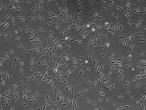
[0045] Example 1: Culture and passage of adipose stem cells

[0046] 1) Adipose tissue was extracted from the abdomen of volunteers, washed 3 times with PBS solution under sterile conditions, washed away red blood cells, and cut into fine particles with a scalpel;

[0047] 2) Add 0.1% collagenase solution 2 times the volume of adipose tissue, shake at 37°C for 2-3 hours, and digest;

[0048] 3) Collect the liquid after shaking and digesting, 1500rpm / 10min, discard the supernatant, and resuspend the pellet with low-sugar DMEM medium containing 10% FBS;

[0049] 4) Filtrate with a 200-mesh fine sieve to remove residual tissue impurities in the suspended cell liquid, and place the filtrate in a centrifuge tube;

[0050] 5) 1500rpm / 10min, discard the supernatant, resuspend the pellet in low-sugar DMEM medium containing 10% FBS, and culture in a 37°C, 5% CO2 incubator. These are primary cells. After 5 days, change the medium for the first time;

[0051] 6) When the confluence of ...

Embodiment 2

[0053] Example 2: Induction of differentiation of adipose stem cells into chondrocytes

[0054] (1) Preheating

[0055] Preheat 1.2% sodium alginate solution (alginate suspension prepared with 0.9% NaCl) and calcium chloride solution to 37°C.

[0056] (2) Preparation of adipose stem cell-sodium alginate suspension

[0057] The adipose stem cells (ADSCs) cultured in the T175 bottle to the P3 generation covered about 90% of the bottom of the bottle. The cells were washed with PBS solution for 3 times, and 2-3ml of 0.25% trypsin digestion solution was added to prepare the ADSCs suspension. The cell concentration was 4.5 ×10 6 a / ml;

[0058] The ADSCs suspension was thoroughly mixed with 1.2% sodium alginate solution at a volume ratio of 3:1 to make a cell concentration of 6.0×10 6 Adipose stem cell-sodium alginate suspension per ml, gently blow with a pipette to prevent the generation of air bubbles.

[0059] (3) Preparation of adipose stem cell-calcium alginate microbeads ...

Embodiment 3

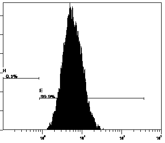
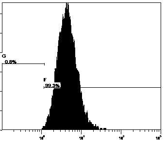
[0073] Embodiment 3: MTT method detects cell proliferation activity

[0074] The adipose stem cells-calcium alginate microbeads of the above four groups on the 3d, 7d, and 14d were respectively inoculated into a 96-well plate, and 100 μl of cell suspension was added to each well, and the cell density was 5×10 4 a / m. There were 3 wells in each group, and a blank control group was set at the same time.

[0075] Add 10 μl of MTT solution (5 mg / ml) to each well, continue to incubate for 4 h in a 37°C, 5% CO2 incubator, carefully discard the supernatant in the well, add 150 μl of dimethyl sulfoxide (DMSO), shake for 10 min, and The absorbance of each well was measured at OD490nm with an immunodetector.

[0076] According to the MTT test, on the 3d, 7d, and 14d after induction, the OD values ​​of the experimental groups B, C, and D were significantly higher than those of the control group A. On the 14th day, the OD value of the control group A was 0.26, and the OD value of the ex...


PUM

PropertyMeasurementUnit
diameteraaaaaaaaaa

Abstract

The invention provides a method for inducing differentiation of adipose derived stem cells into chondrocyte. The method comprises the steps of preparing adipose derived stem cell-sodium alginate suspension, preparing adipose derived stem cell-calcium alginate microbeads, and performing induced culture. According to the method for inducing differentiation of adipose derived stem cells into chondrocyte, an incomplete cartilage forming induced culture solution containing PRP and transforming growth factor-beta superfamily members (TGF-[beta]1, IGF-1 and BMP-6) is utilized, and PRP and transforming growth factor-beta superfamily members are in united use, so that synergistic reactions are achieved, Sox-9 gene expression can be raised, differentiation of induced adipose derived stem cells intohypertrophy phenotypes is restrained, cell proliferation is promoted, and induced differentiation efficiency is improved. The proliferation vitality of the chondrocyte obtained through induced differentiation for 13-15 days by the method disclosed by the invention is 3.4 times of that of a control group A, 2.2 times of that of an experimental group B, and 1.95 times of that of an experimental group C.

Description

technical field [0001] The invention relates to a method for inducing fat stem cells to differentiate into chondrocytes, which belongs to the technical field of stem cells. Background technique [0002] Lesions caused by articular cartilage defects are relatively common in clinical practice, and cartilage trauma is extremely difficult to repair, because there are no blood vessels and nerves in cartilage tissue, and its characteristics determine that cartilage cannot be regenerated. Traditional treatment methods such as joint washing, microcracks, mosaic plastic surgery, etc., can improve the clinical symptoms of patients in the short term, but the long-term effect is not satisfactory; joint prosthesis replacement can eliminate joint pain and restore function, but it is expensive and has many complications; Autologous or allogeneic chondrocytes are difficult to extract, the number of cells is small, and their proliferation ability is limited, which limits their clinical appli...

Claims


Application Information

Patent Timeline
Patent Type & Authority Applications(China)
IPC IPC(8): C12N5/0775C12N5/077C12N11/10C12N11/04
CPCC12N5/0655C12N11/10C12N11/04C12N2506/1384C12N2501/155C12N2501/15C12N2501/105C12N2501/33C12N2500/38C12N2501/39C12N2513/00
Inventor 刘明录卢永灿金海锋王立新强邦明张传鹏冯建海李希鹏韩庆梅许淼
Owner SHANDONG XINRUI BIOTECH CO LTD
Why Eureka
Try Eureka