Exosome detection and typing micro-fluidic chip and exosome detection and typing method

A microfluidic chip and exosome technology, applied in chemical instruments and methods, measurement devices, analytical materials, etc., can solve the problems of low separation efficiency, limited speed and diversity, and long time consumption.

Active Publication Date: 2020-10-20
RUIJIN HOSPITAL AFFILIATED TO SHANGHAI JIAO TONG UNIV SCHOOL OF MEDICINE
View PDF8 Cites 6 Cited by
  • Summary
  • Abstract
  • Description
  • Claims
  • Application Information

AI Technical Summary

Problems solved by technology

The current gold standard methods for exosome capture and identification, such as ultracentrifugation and western blot, suffer from low separation efficiency, low sensitivity, long time-consuming and high sample consumption
Microfluidics has recently been used to exploit exosome analysis, but progress in improving sensitivity, speed, and diversity has been rather limited

Method used


Image

  • Exosome detection and typing micro-fluidic chip and exosome detection and typing method
  • Exosome detection and typing micro-fluidic chip and exosome detection and typing method
  • Exosome detection and typing micro-fluidic chip and exosome detection and typing method

Examples


Embodiment 1

[0037] Embodiment 1 Microfluidic Chip Fabrication Method

[0038] 1. Fabrication of antibody strip chip

[0039] 1. Fabrication of PDMS chips

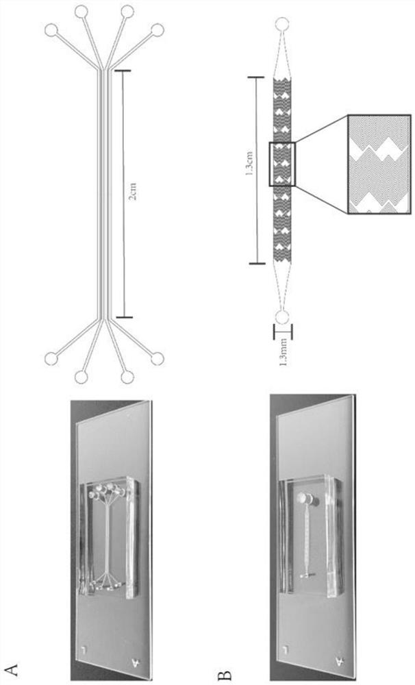
[0040] (1) Use CAD software to draw the graphics of the chip, use a high-resolution laser phototypesetter to draw a photolithography mask on the photographic film, and then spin-coat the SU-8 photoresist layer on the silicon wafer, and perform high-temperature curing treatment on it After covering with a photolithography mask, expose the cured SU-8 photoresist on a photolithography machine, and finally develop it in a developer to obtain a positive silicon wafer template.

[0041] (2) PDMS prepolymer and PDMS curing agent are mixed at a ratio of 10:1, and poured on the prepared positive mold template after pumping in a vacuum, and peeled off after high temperature curing to obtain a chip with a microstructure, as required Punch holes (antibody perfusion holes, antibody outflow holes).

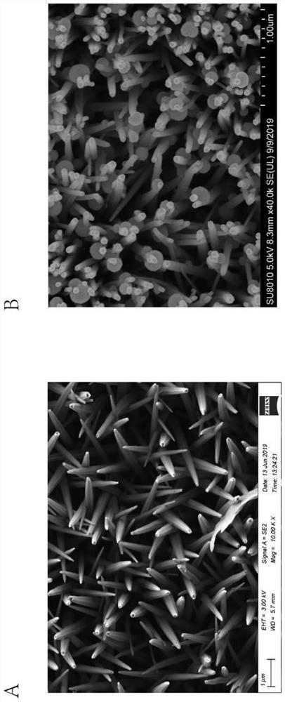
[0042] 2. Preparation of ZnO seed layer on gla...

Embodiment 2

[0080] Example 2 Application of microfluidic chip for detection and typing of exosomes

[0081] 1. Sample injection

[0082] Take 50 μl of HOS cell-derived exosome suspension, add it into the injection hole, the syringe pump draws the sample at a speed of 2 μL / min, and collect the outflowing waste liquid at the sample outlet.

[0083] 2. Rinse the channel with PBS.

[0084] 3. Fluorescent dye perfusion:

[0085] DiO dye is a lipophilic membrane dye. The membrane of exosomes is the same as the cell membrane, which is a phospholipid bilayer, so DiO dye can quickly bind to the membrane surface of exosomes, making the exosomes emit green fluorescence, so that Observe under a fluorescence microscope.

[0086] DiO preparation: Weigh 0.001 g of DiO powder with an analytical balance, dissolve it in 226 μl of DMSO, and prepare a 5 mM DiO stock solution, and store the stock solution in a -20 degree refrigerator. Take 1 μl of DiO dye stock solution in an EP tube, add 99 μl of PBS to ...

Embodiment 3

[0091] Example 3 Optimization of Microfluidic Chip Structure and Detection Conditions

[0092] Some parameters of the structure and detection conditions of the microfluidic chip were changed, and the remaining parameters were the same as in Examples 1 and 2, so as to compare the exosome capture efficiency under different structures and detection conditions.

[0093] 1. Chip injection flow rate screening

[0094] 1. Zinc oxide chip preparation.

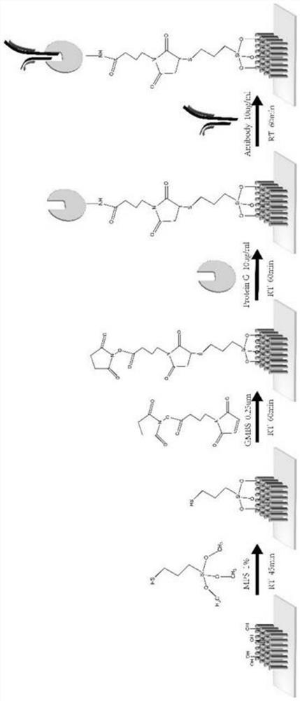
[0095] 2. Zinc oxide surface modified CD63+CD81 antibody.

[0096] 3. Put 6 zinc oxide chips prepared and modified above CD63+CD81 on the table, add 50 μl of HOS cell line exosome suspension respectively, turn on the syringe pump and inject 0, 1, 2, 4 , 8, 10μl / min speed to draw samples.

[0097] 4. After the above process, wash the channel with PBS 3 times.

[0098] 5. Add 50 μM DiO solution and incubate at room temperature for 50 min.

[0099] 6. Rinse with PBS 3 times.

[0100] 7. Observe under a fluorescence microscope and co...


PUM

PropertyMeasurementUnit
heightaaaaaaaaaa
widthaaaaaaaaaa
lengthaaaaaaaaaa

Abstract

The invention relates to an exosome detection and typing micro-fluidic chip and an exosome detection and typing method. The micro-fluidic chip comprises an antibody strip chip and a sample injection chip, CD63 and CD81 antibodies are coupled to the antibody strip chip, and the coupling method comprises the following steps of: firstly, preparing a glass slide with a zinc oxide nano array, then carrying out MPS modification, GMBS modification and G protein modification, and finally connecting the CD63 and CD81 antibodies. A fishbone-shaped microstructure is arranged above a main channel of the sample injection chip, and the height of the channel is 20 microns. The exosome detection and typing method is completed by adopting the micro-fluidic chip, and the sample injection speed is 1.8-2.2 [mu] L / min. The micro-fluidic chip provided by the invention can capture related exosomes to the greatest extent, and is very high in sensitivity.

Description

technical field [0001] The invention belongs to the field of biochips, and in particular relates to a microfluidic chip for detecting and typing exosomes and a method for detecting and typing exosomes. Background technique [0002] Microfluidics integrates the basic operation units of sample preparation, reaction, separation, and detection in biological, chemical, and medical analysis processes into a micron-scale chip to automatically complete the entire analysis process. Due to its great potential in the fields of biology, chemistry, and medicine, it has developed into a new research field interdisciplinary in biology, chemistry, medicine, fluid, electronics, materials, and machinery. The microfluidic chip is the main platform for the realization of microfluidic technology, and its device features are mainly its effective structures (channels, reaction chambers and other functional components) containing fluids that are micron-scale in at least one latitude. Owing to the ...

Claims


Application Information

Patent Timeline
Patent Type & Authority Applications(China)
IPC IPC(8): G01N33/68G01N33/577B01L3/00
CPCG01N33/6872G01N33/577B01L3/502707Y02A50/30
Inventor 沈宇辉许一奇
Owner RUIJIN HOSPITAL AFFILIATED TO SHANGHAI JIAO TONG UNIV SCHOOL OF MEDICINE
Why Eureka
Try Eureka