Application of carbon quantum dots in targeted cell nucleolus wash-free imaging

A technology of carbon quantum dots and cell nucleoli, which is applied in the application field of carbon quantum dots in the no-clean imaging of targeted cell nucleoli, can solve the problems of complex preparation methods, complex operations, and cell damage, and achieve simple preparation and simple operation process , time-saving effect

Active Publication Date: 2021-03-09
SHANXI UNIV
View PDF39 Cites 4 Cited by
  • Summary
  • Abstract
  • Description
  • Claims
  • Application Information

AI Technical Summary

Problems solved by technology

[0005] In order to solve the problems of complex preparation method and operation of materials currently used for cell nucleolus imaging, which require multiple

Method used


Image

  • Application of carbon quantum dots in targeted cell nucleolus wash-free imaging
  • Application of carbon quantum dots in targeted cell nucleolus wash-free imaging
  • Application of carbon quantum dots in targeted cell nucleolus wash-free imaging

Examples


Embodiment 1

[0030] Embodiment 1: Preparation of N-CDs

[0031] Step 1: Accurately weigh 0.05 g of m-phenylenediamine and 0.05 g of p-aminobenzoic acid, completely dissolve them in 10 mL of absolute ethanol, add the resulting solution into a polytetrafluoroethylene liner, and heat at 180°C for 12 h. After the reaction kettle was naturally cooled to room temperature, the brown solution was taken out from the inner tank.

[0032] In step 2, the ethanol solvent in the brown solution was removed by rotary evaporation to obtain a brown viscous substance, which was dissolved by adding secondary water, and centrifuged at 10,000 rpm for 5 min to obtain a brownish-yellow supernatant.

[0033] Step 3, the brown supernatant was treated with a 500-1000 Da dialysis bag for 2 days, during which the water was changed every 6 h, and the dialyzed solution was freeze-dried to obtain a brown N-CDs solid powder.

[0034] Step 4: Weigh 0.01 g of N-CDs solid powder into a beaker, add 10 mL of secondary water t...

Embodiment 2

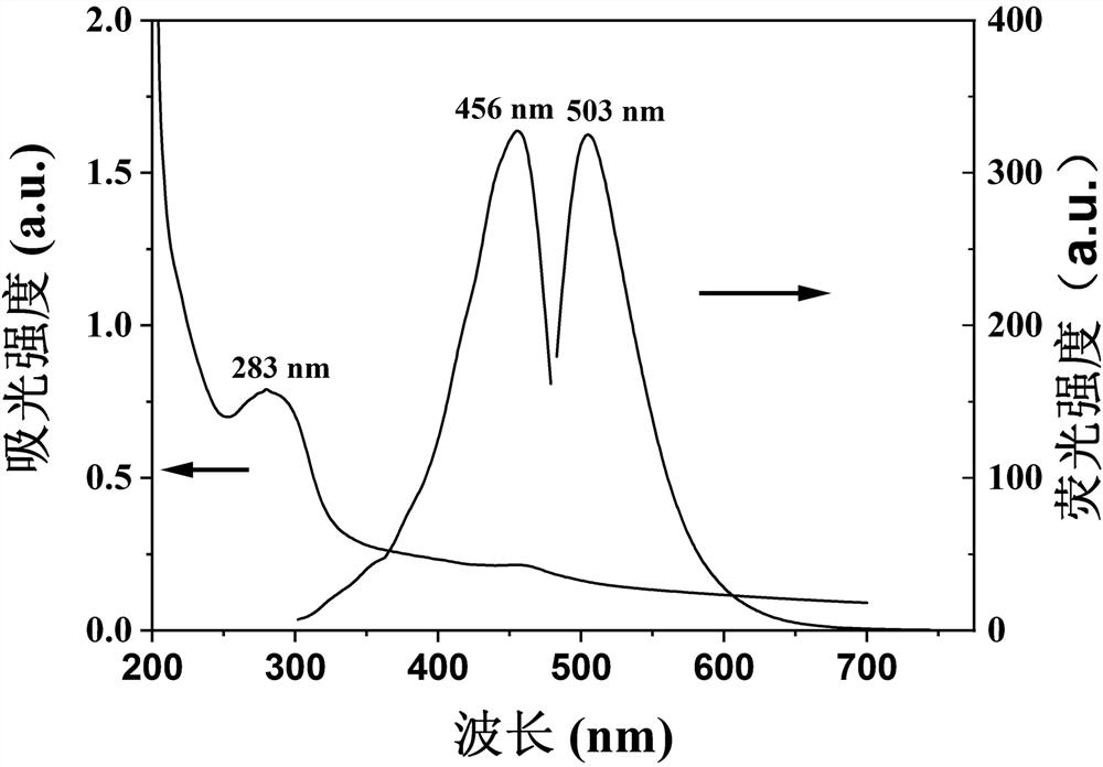
[0035] Example 2: Characterization of N-CDs: See Figure 1-6 . figure 1 In the ultraviolet absorption spectrum of N-CDs, there is a strong absorption peak at 283 nm, which is formed by n→π of C=O * caused by the transition; figure 1 The optimal excitation and emission peaks of the N-CDs are located at 456 nm and 503 nm, respectively.

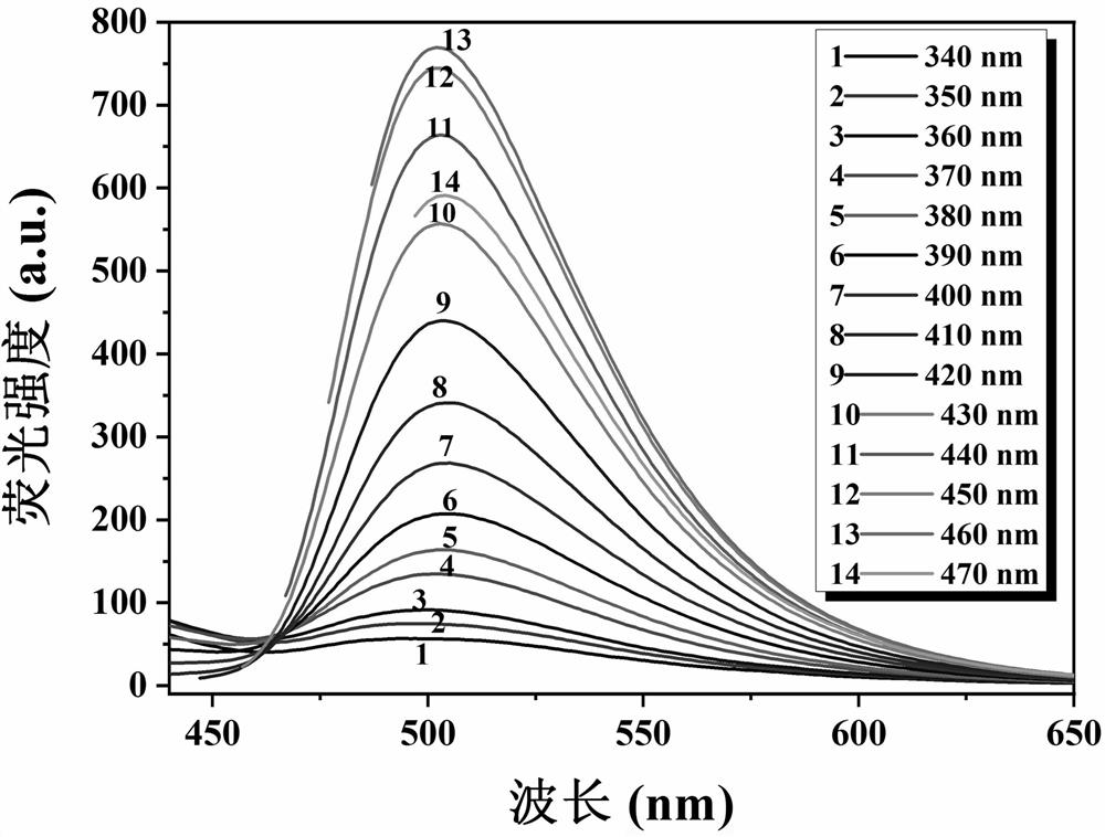
[0036] figure 2 It is the emission spectrum of N-CDs at different excitation wavelengths. When the excitation wavelength changes from 340 nm to 470 nm, the emission wavelength does not shift, indicating that N-CDs have excitation wavelength independence.

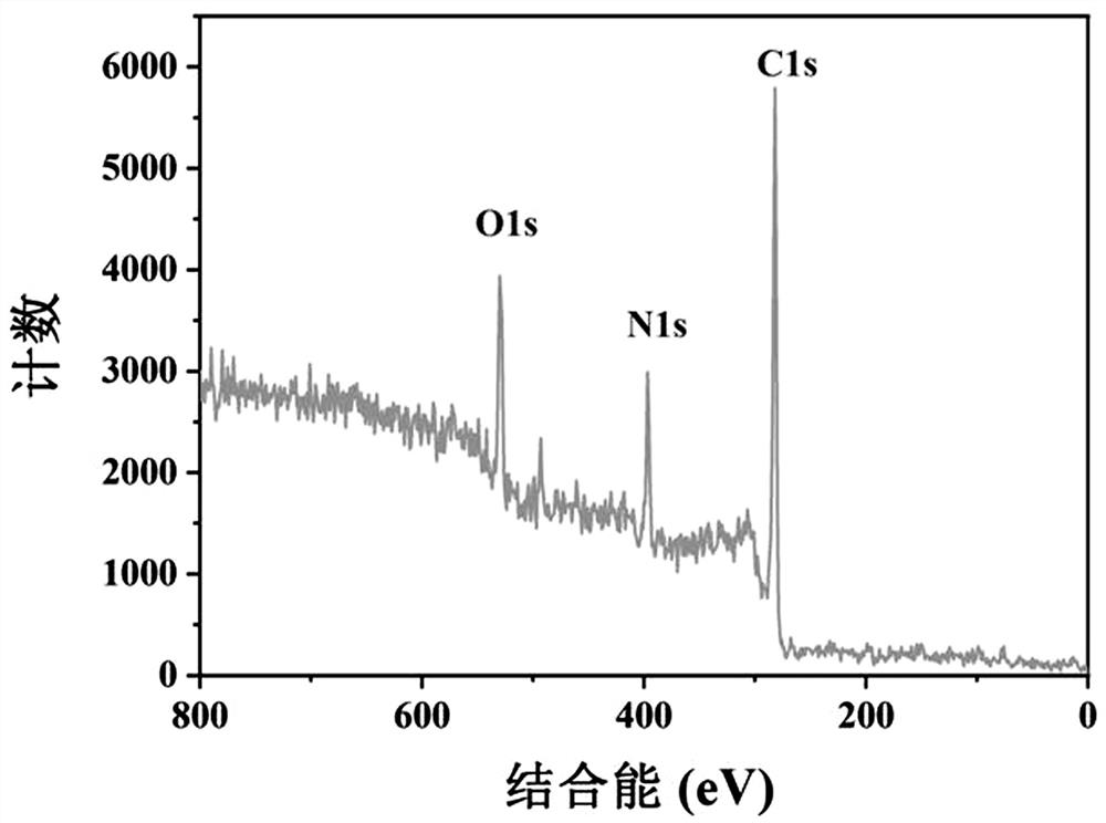
[0037] image 3 It is the X-ray photoelectron spectrum of N-CDs, which shows that N-CDs are mainly composed of three elements: C, N, and O.

[0038] Figure 4 It is the infrared spectrum of N-CDs, which shows that N-CDs are rich in functional groups such as O-H / N-H, C-H, C=N, C-N, C-O, etc., ensuring that N-CDs have good water solubility, and N-CDs have positive potential (25.2 mV ), s...

Embodiment 3

[0041] Example 3: Toxicity test of N-CDs on HeLa cells

[0042] HeLa cells were grown in DMEM medium enriched with 10% fetal bovine serum, 100 U / mL penicillin, and 100 μg / mL streptomycin at 37°C, 5% CO 2 cultivated in the environment. Cells in the logarithmic growth phase were divided into 8×10 4 / mL was inoculated into 96-well culture plates (100 μL culture solution per well), and incubated at 37°C, 5% CO 2 Incubate in the environment for 24 h. After the cells were completely adhered to the wall, the cells were divided into a blank control group and an experimental group. There were 6 replicate wells in each group. The final concentrations of N-CDs added to the experimental group were 0.05, 0.5, 5, 25, and 50 µg / mL, respectively. . After the cells were further incubated for 24 h, the liquid in the wells was aspirated, and 100 μL of DMEM medium containing MTT (0.5 mg / mL) was added to each well. After the cells were further incubated for 4 h, the supernatant was aspirated,...


PUM

PropertyMeasurementUnit
Particle sizeaaaaaaaaaa

Abstract

The invention belongs to the technical field of fluorescence imaging, and provides application of carbon quantum dots in targeted cell nucleolus wash-free imaging. A preparation method comprises the following steps: by taking m-phenylenediamine and p-aminobenzoic acid as a carbon source and a nitrogen source, preparing brown-yellow carbon quantum dots (N-CDs) by a one-step hydrothermal method, andremoving an ethanol solvent by rotary evaporation to obtain a brown viscous material; dissolving the obtained brown viscous material in secondary water, centrifuging to remove insoluble substances, dialyzing to remove unreacted reaction precursor small molecules, and freeze-drying to obtain brown N-CDs solid powder, namely the carbon quantum dots for targeted cell nuclei wash-free imaging. High-resolution cell imaging images of HeLa and PC12 are obtained by laser confocal imaging, which shows that N-CDs can successfully target cell nucleolus for no-clean imaging, and the reagent is an excellent cell nucleolus imaging reagent. The preparation method is simple, can target cell nuclei without modification, has low requirements on instruments and equipment, is simple and convenient to operate, does not need to wash nuclei for imaging, saves time and can avoid cell damage.

Description

technical field [0001] The invention belongs to the technical field of fluorescence imaging, and in particular relates to the application of carbon quantum dots in targeted cell nucleolus wash-free imaging. Background technique [0002] The nucleolus is the most important subnuclear structure in the cell, and its main function is to synthesize ribosomes and rRNA. The size and shape of the nucleolus vary with the species of organisms, cell types and cell metabolism. Studies have shown that changes in the number or shape of nucleoli may be related to cancer and can be used as targets for diagnosis and treatment of cancer, so understanding the state of nucleoli is the key to diagnosis and treatment of cancer. [0003] Materials currently used for cell nucleolus imaging mainly include: gold nanoclusters, metal chelates, small organic molecules, coordinated quantum dots, and graphene. These materials can be successfully located in the nucleolus of cells, but their preparation p...

Claims


Application Information

Patent Timeline
IPC IPC(8): G01N21/64C09K11/65C09K11/08B82Y20/00B82Y30/00B82Y40/00C01B21/06
CPCB82Y20/00B82Y30/00B82Y40/00C01B21/0605C09K11/0883C09K11/65G01N21/6486
Inventor 弓晓娟张俐王旭董文娟刘洋董川
Owner SHANXI UNIV
Why Eureka
Try Eureka