
Marker molecules at different time points in endometrial secretion period and screening method thereof

A technology of endometrium and secretory phase, which is applied in the biological field, can solve the problem of not being able to know the molecular changes of the endometrium in the secretory phase, so as to reduce the burden on patients and social pressure, and improve the success rate

Pending Publication Date: 2021-08-24
NANJING DRUM TOWER HOSPITAL
View PDF3 Cites 9 Cited by
  • Summary
  • Abstract
  • Description
  • Claims
  • Application Information

AI Technical Summary

Problems solved by technology

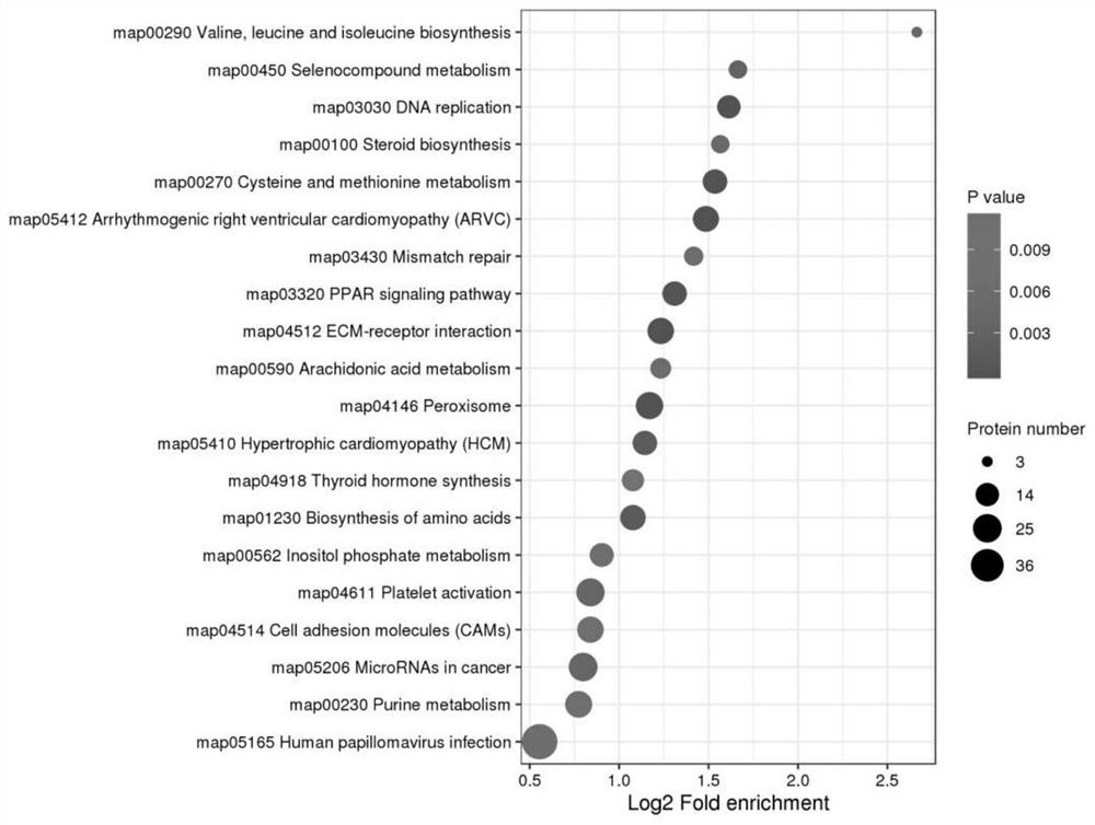
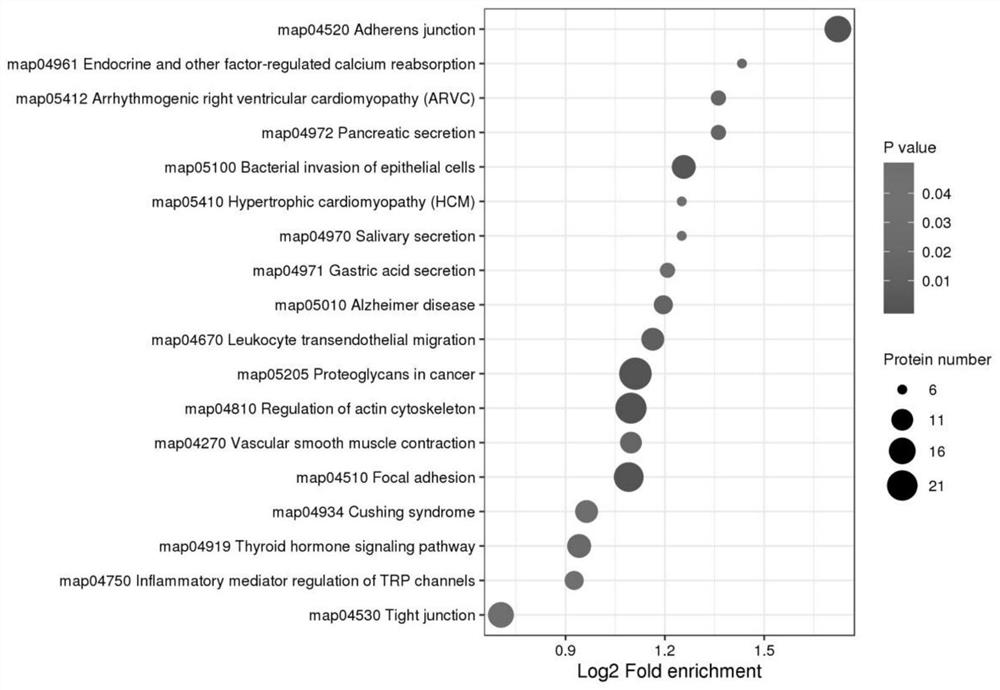
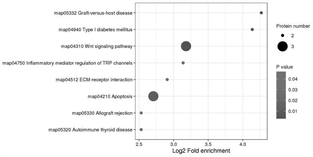
However, this method cannot obtain the molecular changes of the endometrium during the secretory phase. This method only has one time and only the proteome level. This application includes 6 time points from early secretory to late secretory, and includes proteomics and phosphorylation Two Dimensions of Modified Omics

Method used


Image

  • Marker molecules at different time points in endometrial secretion period and screening method thereof
  • Marker molecules at different time points in endometrial secretion period and screening method thereof
  • Marker molecules at different time points in endometrial secretion period and screening method thereof

Examples


Embodiment 1

[0089] Example 1 protein extraction

[0090] Take the sample out from -80°C, weigh an appropriate amount of tissue sample into a mortar pre-cooled with liquid nitrogen, add liquid nitrogen to fully grind to powder, and add 4 times the volume of sample lysis buffer (1% Triton X-100, 1% protease inhibitor, 1% phosphatase inhibitor) for ultrasonic lysis; 4°C, 12000g centrifugation for 10 min to remove cell debris, transfer the supernatant to a new centrifuge tube, and use the BCA kit for protein concentration determination.

Embodiment 2

[0091] Example 2 trypsin hydrolysis

[0092] Take an equal amount of protein from each sample in Example 1 for enzymatic hydrolysis, adjust the volume to the same volume with the lysate, slowly add 20% TCA at a final concentration, vortex and mix, precipitate at 4°C for 2 hours, centrifuge at 4500g for 5 minutes, and discard the supernatant , wash the precipitate with pre-cooled acetone for 2-3 times, dry the precipitate, add TEAB with a final concentration of 200mM, ultrasonically break up the precipitate, add trypsin at a ratio of 1:50 (protease: protein, m / m), and enzymolyze Overnight, add dithiothreitol (DTT) to make the final concentration 5mM, reduce at 56°C for 30min, then add iodoacetamide (IAA) to make the final concentration 11mM, and incubate at room temperature for 15min in the dark.

Embodiment 3

[0093] Example 3 For phosphorylation modification proteomics, modification enrichment

[0094] Dissolve the peptides obtained in step (2) in the enrichment buffer solution (50% acetonitrile / 0.5% acetic acid), transfer the supernatant to the pre-washed IMAC material, place it on a rotary shaker and incubate with gentle shaking, After the incubation, wash the material three times with buffer solution 50% acetonitrile / 0.5% acetic acid and 30% acetonitrile / 0.1% trifluoroacetic acid in sequence, and finally use 10% ammonia water to elute the phosphopeptide, collect the eluate and dry it by vacuum freeze, After drying, desalt according to the instructions of C18ZipTips, vacuum freeze and dry for liquid mass spectrometry analysis.


PUM


Abstract

The invention discloses a protein extraction and treatment process at different time points (including early, middle and late periods of secretion: LH_2, LH_5, LH_7, LH_9, LH_10 and LH_12, and LH_0 on the day of injection of hCG) in a human endometrial tissue secretion period, and a protein spectrum and a phosphorylation modification mass spectrum are carried out to obtain molecular expression modes at different time points in the secretion period. The molecular expression modes can provide a basis for further knowing the dynamic change of the human endometrial tissue secretion period, and is beneficial to judging an endometrial implantation window period and guiding the embryo transplantation time in an assisted reproduction technology.

Description

technical field [0001] The invention belongs to the field of biotechnology, and in particular relates to molecular changes at different time points in endometrium secretory phase and a screening method thereof. Background technique [0002] Reproductive health is not only related to the quantity and quality of the birth population, but also related to the national economy and people's livelihood. According to the statistics of the World Health Organization, the reproductive problems of the global population of childbearing age are becoming increasingly prominent. Although the emergence of assisted reproductive technology has alleviated the problem of reduced human fertility to a certain extent, the success rate of in vitro fertilization-embryo transfer has been hovering around 50% worldwide, and the failure of embryo implantation is the bottleneck factor. [0003] A good endometrium is a prerequisite for successful embryo implantation. The mid-secretory endometrium in a rec...

Claims


Application Information

Patent Timeline
IPC IPC(8): G01N30/88G01N30/02G01N30/72
CPCG01N30/88G01N30/02G01N30/72G01N2030/8831
Inventor 颜桂军孙海翔刘洋曹志文李东
Owner NANJING DRUM TOWER HOSPITAL
Learn More