Preparation method of exosome bionic preparation for synergistically promoting wound healing and preparation thereof

A wound healing and exosome technology, applied in the field of cell biology, can solve the problems of difficulty in exerting the best biological activity, poor drug loading, and low yield, and achieve the effects of good application prospects, low cost and high safety.

Active Publication Date: 2021-11-02
苏州尼达生物科技有限公司
View PDF7 Cites 0 Cited by
  • Summary
  • Abstract
  • Description
  • Claims
  • Application Information

AI Technical Summary

Problems solved by technology

[0003] Exosomes derived from umbilical cord mesenchymal stem cells have become a research hotspot in wound healing in recent years. The CD73, CD90, CD105 and other cytokines expressed on the surface can not only locate the injury and inflammation site, but also release paracrine factors such as tissue Repair factors, angiopoietin, and immunomodulatory factors promote wound healing, participate in a series of important processes such as inflammation regulation, angiogenesis, and epithelial collagen deposition in the wound healing process, and are non-immunogenic. It is difficult to exert the best biological activity due to the influence of oxygen environment, and its low yield and poor drug loading further limit its application.

Method used


Image

  • Preparation method of exosome bionic preparation for synergistically promoting wound healing and preparation thereof
  • Preparation method of exosome bionic preparation for synergistically promoting wound healing and preparation thereof
  • Preparation method of exosome bionic preparation for synergistically promoting wound healing and preparation thereof

Examples


Embodiment 1

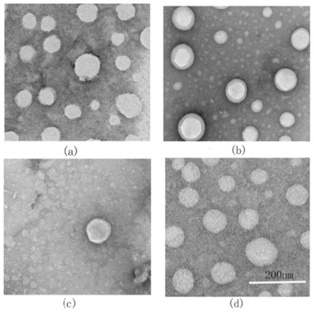
[0046] (1) Preparation of catalase-photosensitizer micelles (CAT-Ce6-m): weigh Ce6 (60 mg) and dissolve in 10 mL DMSO, add EDCI (100 mg) and NHS (150 mg), and stir at room temperature for 6 h in the dark, Add catalase CAT (20 mg), stir at room temperature for 24 hours in the dark, dialyze the reaction solution with a dialysis bag with a cutoff of 3.5 kD for 24 hours at room temperature to obtain a micellar solution, and place the dialyzed micellar solution in a vacuum freezer Freeze-dry in a dryer to obtain CAT-Ce6-m powder.

[0047] (2) Preparation of micellar liposomes (CAT-Ce6-m@lip) loaded with catalase-photosensitizer: take soybean lecithin (223mg) and cholesterol (28mg), dissolve them in 5mL absolute ethanol, and keep at 25°C Form a film under reduced pressure for 15 minutes, dry with nitrogen to remove the organic solvent, add 5 mL of PBS solution (pH 7.4) containing 2 mg / mL of catalase-photosensitizer micelles (CAT-Ce6-m), and hydrate at 25 °C for 30 min. Ultrasound w...

Embodiment 2

[0054] (1) Preparation of catalase-photosensitizer micelles (CAT-Ce6-m): same as Example 1;

[0055] (2) Preparation of micellar liposomes (CAT-Ce6-m@lip) loaded with catalase-photosensitizer: take soybean lecithin (200mg) and cholesterol (50mg), dissolve them in 5mL absolute ethanol, and keep at 25°C The film was formed under reduced pressure for 15 minutes, and dried with nitrogen to remove the organic solvent. Add 5 mL of PBS solution (pH 7.4) containing 2 mg / mL of catalase-photosensitizer micelles (CAT-Ce6-m), hydrate at 25 °C for 30 min, sonicate the probe at 100 W for 10 min, and store at 4 °C for later use;

[0056] (3) Extraction and separation of exosomes from mesenchymal stem cells, same as in Example 1;

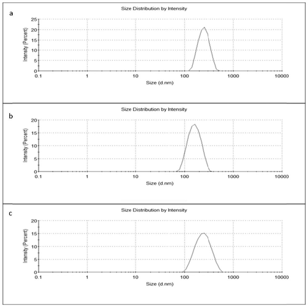
[0057] (4) Mix the liposome solution obtained in step (2) with the exosome solution obtained in step (3) in a mass ratio of 2:1, and pass through 5 μm, 1 μm, 500 nm and 200 nm polycarbonate in sequence with an extruder membrane, repeatedly extruded 10 times and t...

Embodiment 3

[0061] (1) Preparation of catalase-photosensitizer micelles (CAT-Ce6-m): same as Example 1;

[0062] (2) Preparation of micellar liposomes (CAT-Ce6-m@lip) loaded with catalase-photosensitizer: take soybean lecithin (160mg) and cholesterol (40mg), dissolve them in 5mL absolute ethanol, and keep at 25°C The film was formed under reduced pressure for 15 minutes, and dried with nitrogen to remove the organic solvent. Add 5 mL of PBS solution (pH 7.4) containing 2 mg / mL of catalase-photosensitizer micelles (CAT-Ce6-m), hydrate at 25 °C for 30 min, sonicate the probe at 100 W for 10 min, and store at 4 °C for later use;

[0063] (3) Extraction and separation of exosomes from mesenchymal stem cells, same as in Example 1;

[0064] (4) Mix the liposome solution obtained in step (2) with the exosome solution obtained in step (3) in a mass ratio of 1:5, and pass through 5 μm, 1 μm, 500 nm and 200 nm polycarbonate in sequence with an extruder membrane, repeatedly extruded 10 times and t...


PUM

PropertyMeasurementUnit
diameteraaaaaaaaaa

Abstract

The invention discloses a preparation method of an exosome bionic preparation for synergistically promoting wound healing and a preparation thereof. The preparation method of the preparation comprises the following steps: preparing catalase-photosensitizer micelle, preparing ROS response lipidosome carrying catalase-photosensitizer micelle, extracting, separating and purifying exosome, mixing the lipidosome with the exosome, mediating membrane fusion between the liposome and the exosome by an extrusion method, and mixing the membrane fusion carrier and gel, and uniformly stirring the mixture to prepare the preparation. According to the preparation prepared by the invention, photodynamic therapy and biological therapy are combined, catalase-photosensitizer micelles are wrapped by a membrane fusion carrier, accumulated hydrogen peroxide is eliminated on a wound surface, oxygen is supplemented, and the photosensitizer generates controllable singlet oxygen to resist inflammation and sterilize, so that favorable conditions are created for treating the wound surface by the exosome; meanwhile, the gel provides a favorable moist environment for wound healing, and the gel is high in safety, good in stability, convenient to apply, simple in processing technology, low in cost and good in application prospect.

Description

technical field [0001] The invention belongs to the field of cell biology, and in particular relates to a preparation method of an exosome biomimetic preparation (CAT-Ce6-m@lip-exo-gel) that synergistically promotes wound healing and its preparation. Background technique [0002] Wound healing depends on many physiological processes, including cell migration (transfer of inflammatory cells to the injury site), cell proliferation (angioblasts and fibroblasts), apoptosis, and reconstruction of the dermis through the synthesis of extracellular matrix (ECM) proteins, when Inflammatory cells produce excess reactive oxygen species (ROS) such as superoxide anion, H 2 o 2 Time, singlet oxygen, etc. cause tissue necrosis, vascular dysfunction, etc., making it difficult for wounds to heal, and wounds that do not heal for a long time will cause other infections, entering a vicious circle. A good wound healing process needs to inhibit or kill the pathogenic bacteria that cause severe ...

Claims


Application Information

Patent Timeline
Patent Type & Authority Applications(China)
IPC IPC(8): A61K41/00A61K38/44A61K35/28A61K9/107A61K9/127A61K9/06A61K47/24A61K47/28A61K47/32A61P17/02A61P31/04C12N5/0775
CPCA61K41/0071A61K38/44A61K35/28A61K9/1075A61K9/127A61K9/06A61K47/24A61K47/28A61K47/32A61P17/02A61P31/04C12N5/0668C12Y111/01006A61K2300/00
Inventor 李斯文戴宇李钰
Owner 苏州尼达生物科技有限公司
Why Eureka
Try Eureka