Drug screening method and three-dimensional tumor slice model culture method

A screening method and three-dimensional culture technology, applied in the field of biomedicine, can solve the problems of inability to obtain cell or tissue information at the molecular level, time-consuming, unfavorable treatment and recovery of patients, and achieve the effect of accelerating precise anti-cancer treatment

Pending Publication Date: 2021-12-24
UNIVERSITY OF MACAU +1
View PDF2 Cites 8 Cited by
  • Summary
  • Abstract
  • Description
  • Claims
  • Application Information

AI Technical Summary

Problems solved by technology

However, this method has certain limitations. The amount of serum or body fluid that the patient can provide is limited, and excessive extraction of the patient's serum or body fluid will cause a certain degree of physical and mental damage to the patient, which is not conducive to the treatment and recovery of the patient.
In addition, this method is also based on the observation of the state of 3D cultured cells or tissues, and cannot obtain cell or tissue information more intuitively from the molecular level. Further analysis and identification can only be carried out after the experiment is over.
[0006] Despite many efforts on how to realistically mimic the tumor microenvironment of cancer patients, challenges remain in creating an in vitro environment applicable to all types of tumors from different individuals
Three-dimensional cell culture models and organoid culture models still have the shortcomings of insufficient sensitivity and reactivity for drug therapy and new drug development, and the drug screening process takes a lot of time, and real-time evaluation of drugs cannot be achieved in a short time and filter

Method used


Image

  • Drug screening method and three-dimensional tumor slice model culture method
  • Drug screening method and three-dimensional tumor slice model culture method
  • Drug screening method and three-dimensional tumor slice model culture method

Examples


preparation example Construction

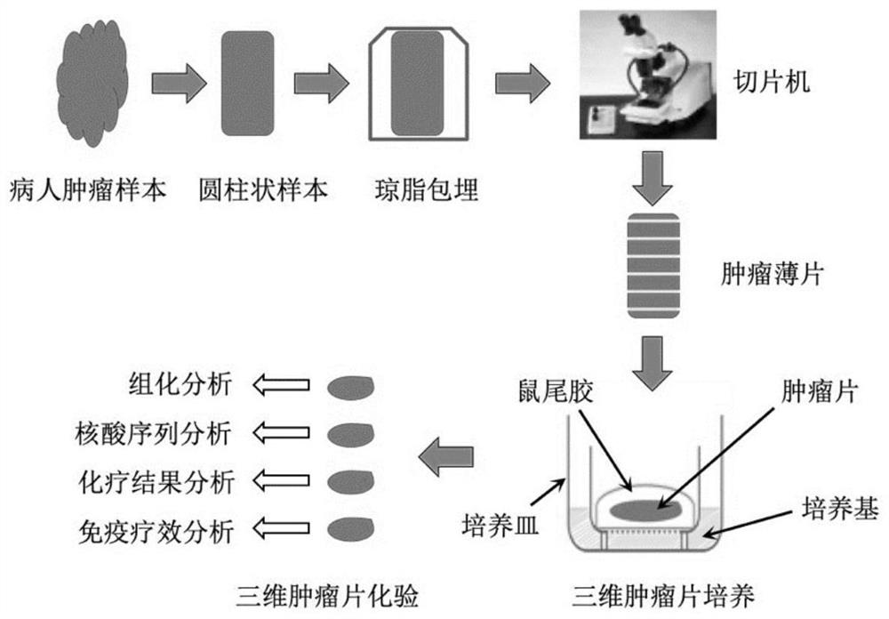
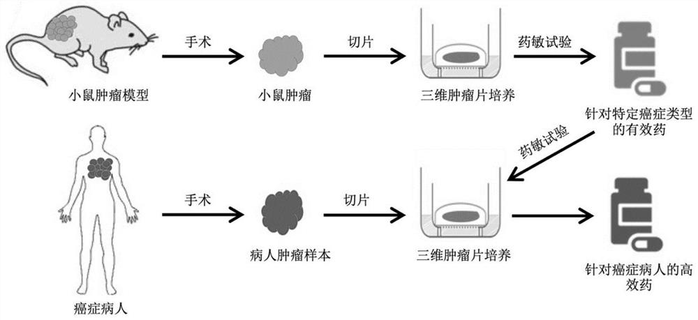
[0028] An embodiment of the present invention provides a drug screening method, which includes the following steps: performing three-dimensional culture on the three-dimensional tumor slice model; wherein, the preparation method of the three-dimensional tumor slice model includes: the tumor labeled with the apoptosis reporter substance The tissue is sliced ​​to obtain the three-dimensional tumor slice model; then, the three-dimensional tumor slice model in three-dimensional culture is processed with candidate drugs, and the candidate drugs are screened based on the signal of the apoptosis reporter substance, which can be referred to figure 1 .

[0029] In this paper, "treating the three-dimensional tumor slice model in three-dimensional culture with a candidate drug" may refer to: contacting the drug with the three-dimensional tumor slice model, specifically by adding the drug to the three-dimensional culture medium. The three-dimensional tumor slice model established by the i...

Embodiment 1

[0059] Three-dimensional tumor slice models are able to maintain the cellular repertoire and immune components of their original tumors.

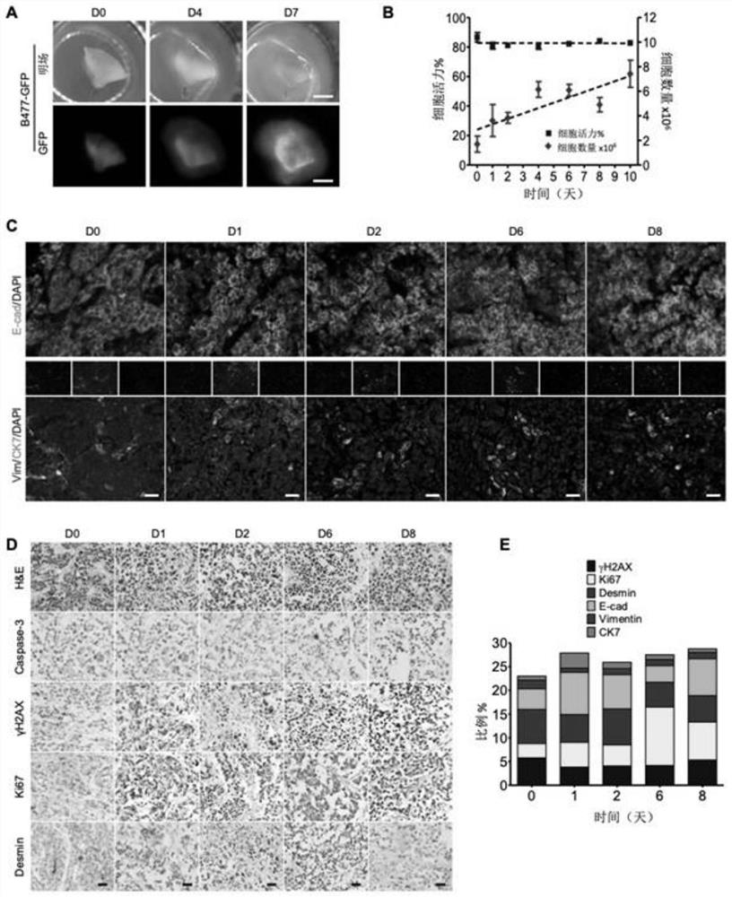
[0060] Three-dimensional tumor slice models were prepared as follows: Fresh tumors were embedded in low-melting point agarose and sliced ​​at a thickness of 300 μm with a vibrating microtome. Then, tumor slices were placed on an air-liquid interface system for culture, and time-course images of the slices were captured from the original tumor before culture (referred to as day 0, D0) to D 7 in culture, using a Leica M165FC fluorescence stereomicroscope .

[0061] Among them, the culture conditions of three-dimensional tumor slices are as follows: A (rat tail collagen I), B (10X Ham's F-12) and C (sterile buffer, 2.2g NaHCO 3 In 100ml0.05N NaOH and 200mM HEPES) according to the ratio of 8:1:1 preparation. Take the prepared 100 μL gel solution and spread it on the membrane (membrane pore size 0.4mm) in the Millicell insert cell culture inne...

Embodiment 2

[0072] Based on FRET time-lapse technology and MTT endpoint method to predict the efficacy of chemical drugs in the three-dimensional tumor slice platform.

[0073] Due to the lack of fluorescent markers in clinical cancer samples, this example uses GFP-labeled cells, C3-labeled cells and non-fluorescent-labeled cells in three-dimensional tumor slices to test the drug response. First, cell viability of fluorescent samples was measured in cisplatin-treated B477-GFP breast tumor sections ( Figure 6 Middle A-B). An increase in GFP signal was found within 7 days during the three-dimensional tumor slice, and this increase was prevented after cisplatin treatment, such as tumor slice size, GFP intensity, etc. These phenomena were consistent with the cell viability determined by the MTT assay.

[0074] MDA-MB-231-C3 tumor slices were next treated with 1 μM bortezomib (drug), and drug responses were measured in a time series study. found that the FRET ratio decreased in a stepwise f...


PUM

PropertyMeasurementUnit
thicknessaaaaaaaaaa

Abstract

The invention discloses a drug screening method and a three-dimensional tumor slice model culture method, and relates to the field of biological medicine. The three-dimensional tumor slice model culture method is established, high-throughput screening of drugs is achieved by combining a label-free technology and/or a cell apoptosis report substance delayed imaging method, and efficient drugs for specific cancer samples are obtained within one week. Immune components of original tumors are completely reserved in three-dimensional tumor slice model culture, and which is possible that an immune checkpoint blocking test is successfully achieved through an immune checkpoint inhibitor. By means of the technology, a cheap, rapid and simple platform is provided for anti-cancer drug discovery, and accurate anti-cancer treatment is accelerated.

Description

technical field [0001] The invention relates to the technical field of biomedicine, in particular to a method for screening drugs and a method for culturing three-dimensional tumor slice models. Background technique [0002] Precision medicine is an approach to tailor interventions that take into account the influence of an individual's genetics, environment, and lifestyle exposures. In the past, drug discovery models used in personalized medicine for cancer treatment have many defects. Human cell lines cultured in 2D can easily lose their original functions, and their morphology, biological function, genetics, and other aspects are very different from human physiology. And dye labeling ignores cellular dynamics and metabolic changes in the drug-treated microenvironment, making it difficult to quantitatively assess drug sensitivity and efficiency in tumor fragments. Therefore, many biological tests to assess the safety and efficacy of drug candidates must be performed in an...

Claims


Application Information

Patent Timeline
Patent Type & Authority Applications(China)
IPC IPC(8): C12Q1/02C12N5/09
CPCG01N33/5011G01N33/5088C12N5/0693C12N2513/00C12N2503/02G01N2500/10
Inventor 邓初夏刘子铭邢富强王冠宇
Owner UNIVERSITY OF MACAU
Why Eureka
Try Eureka