Single-cell protein digital imaging detection method

A single-cell protein and imaging detection technology, applied in the field of protein detection, can solve the problems of large antigen loss, easy signal overlap, multiple washing and staining, etc., achieve good functional stability, no significant change in signal, and save experimental time.

Pending Publication Date: 2021-12-31
SHANGHAI JIAO TONG UNIV
View PDF4 Cites 0 Cited by
  • Summary
  • Abstract
  • Description
  • Claims
  • Application Information

AI Technical Summary

Problems solved by technology

Single-cell western blot detection uses traditional fluorescent labels, with fewer channels, signals are easy to overlap, and detection requires multiple washings, resulting in a large loss of antigens

Method used


Image

  • Single-cell protein digital imaging detection method
  • Single-cell protein digital imaging detection method
  • Single-cell protein digital imaging detection method

Examples


Embodiment 1

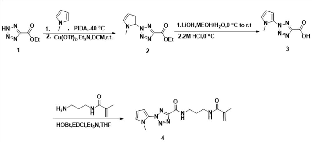
[0131] N-(3-methacrylamidopropyl)-2-(1-methyl-1H-pyrrol-2-yl)-2H-tetrazole-5-formamide is used in preferred embodiments of the present invention, That is, MAP-mPyTC is used as an additive in the protein immobilization gel, refer to the attached figure 2 , the synthesis steps of MAP-mPyTC are as follows:

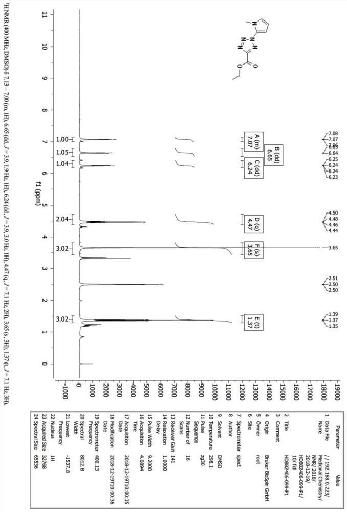
[0132] Step 2.1, preparation of ethyl 2-(1-methyl-1H-pyrrol-2-yl)-2H-tetrazole-5-carboxylate:

[0133] Step 2.1.1, dissolving 5 g, namely 12.5 mmol, of methyl-1 hydrogen-pyrrole in 30 mL of trifluoroethanol;

[0134] Step 2.1.2. At -40°C, add 20 g, i.e. 62.5 mmol, of iodobenzene diacetate to the solution in step 2.1.1, and stir at -40°C for 2 hours under nitrogen protection;

[0135] Step 2.1.3, concentrate the product of step 2.1.2 to black oil, dissolve in dichloromethane;

[0136] Step 2.1.4, add 3g namely 21.6mmol ethyl 5-formate tetrazolium, 3.1g namely 8.6mmol trifluoromethanesulfonate copper (II) and 16ml namely 108mmol triethylamine in step 2.1.3, in nitrogen Und...

Embodiment 2

[0160] In addition, the acrylamide gel based on tetrazolium as the photosensitive group can be substituted, and the photosensitizer in the photosensitive protein immobilized gel can also use other analogs, the general structural formula is shown in formula IV, wherein the R group is Electron-withdrawing group:

[0161]

[0162] Preferably, the R group is selected from one of the following groups.

[0163]

[0164] In step 3-4, it is necessary to prepare a light-sensitive protein immobilization gel, and prepare a single-cell Western blot chip with the chip template in step 1. The schematic diagram of the chip template and the chip in one embodiment of the present invention is as follows Figure 7 As shown, due to the special design of the film mask in step 1, the protruding structure on the surface of the chip template is formed, so that the chip made in step 3 forms a complementary concave structure under the action of the template, that is, micropores . When the loade...

Embodiment 3

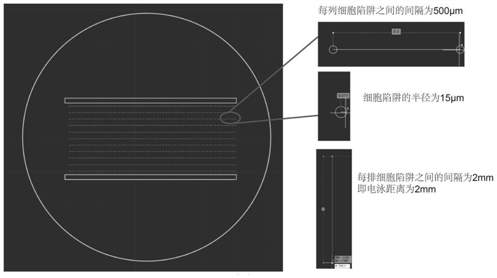
[0166] Such as Figure 7As shown, in a preferred embodiment of the present invention, the western blotting chip prepared in step 3 is a light-sensitive protein immobilized gel attached to one side of the glass slide, and the chip template made in step 1 has multiple Two protruding cylinders form a cylinder array, so that the gel part on the Western blot chip prepared in step 3 has multiple micropores to form a porous microarray. In addition, there are multiple hydrophobic regions on the chip template, composed of hydrophobic The hydrophobic pen is a commonly used tool in immunohistochemistry. It can draw a thick line on the glass. This line is a hydrophobic chemical substance. When the aqueous solution encounters this line, it will be blocked and will not pass through. boundary, so that the gel part of the western blot chip forms a plurality of isolation regions without gel, the diameter of the cylinder is 30 μm, and the width of the isolation regions is 2 mm, and the isolatio...


PUM

PropertyMeasurementUnit
strengthaaaaaaaaaa
diameteraaaaaaaaaa
concentrationaaaaaaaaaa

Abstract

The invention discloses a single-cell protein digital imaging detection method, and relates to the field of protein detection. The single-cell protein digital imaging detection method comprises the steps that a designed film mask is used for preparing a chip template, a protein immobilized gel photosensitizer is synthesized, a western blot chip with light-sensitive protein immobilized gel is prepared in cooperation with the chip template, and after a sample is added, electrophoresis is carried out according to a single-cell immunoblotting (scWB) program, the protein is immobilized through UV crosslinking, target protein immunoblotting detection is carried out by using a labeled antibody, and an analysis result is observed. The invention provides a single-cell protein detection means which is high in throughput, short in time, capable of stably storing data and digitally processing detection results, portable, small and exquisite, stable in function and low in cost.

Description

technical field [0001] The invention relates to the field of protein detection, in particular to a single-cell protein digital imaging detection method. Background technique [0002] Protein analysis of rare cells has become an increasing focus of research. However, limited by sampling technology, enrichment technology or the extremely low content of this type of cell in the body itself, it is difficult to perform systematic protein detection with existing technical means. At present, the most studied rare cells are circulating tumor cells, fertilized eggs, and neuronal cells. They play an important role in disease, development, and early cognitive formation respectively. If dozens or even hundreds of protein levels in rare cells can be analyzed Delineation plays an important role in promoting developmental biology, tumor immunity and other fields. [0003] The existing protein detection technology can be divided into two categories: non-labeled detection and labeled detec...

Claims


Application Information

Patent Timeline
Patent Type & Authority Applications(China)
IPC IPC(8): G01N33/68G01N33/532G01N27/447G01N21/64
CPCG01N33/68G01N33/532G01N27/447G01N21/6458G01N27/62
Inventor 丁显廷谢海洋李山鹤丁屹朱大为
Owner SHANGHAI JIAO TONG UNIV
Why Eureka
Try Eureka