Application of selective autophagy activator QX77 in preparation of medicine for intervening or treating diabetic retinopathy
A diabetic retinal, selective technology for drug combinations, metabolic diseases, sensory diseases, etc.
- Summary
- Abstract
- Description
- Claims
- Application Information
AI Technical Summary
Problems solved by technology
Method used
Image

Examples
Embodiment 1
[0028] Example 1: Diabetic Rat Model Construction and Vitreous Drug Injection
[0029] 1.1 Rat preparation: 4 W SD rats with a weight of about 140 g were purchased from Slack Company and kept in the SPF room of Tongji University Animal Center. Rats were randomly divided into three groups, normal control group (NC), diabetic group (DM), diabetic retinal injection group (DM+QX77).
[0030] 1.2 Modeling by intraperitoneal injection: the rats were starved for 24 hours before the experiment. A single intraperitoneal injection of STZ (60 mg / kg body weight) was used to induce diabetes, and the normal control group was intraperitoneally injected with an equal volume of citric acid solution; 24 hours later, blood was taken from the tail to measure blood sugar, and rats with a blood sugar value higher than 250 mg / dL were used for the test. For intravitreal injection, rats below 250 mg / dL will be excluded.
[0031] 1.3 Intravitreal injection: Rats were anesthetized by intraperitoneal i...
Embodiment 2
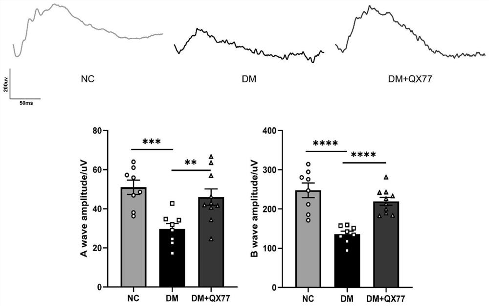
[0032] Embodiment 2: SD rat electroretinogram ERG detects physiological function
[0033] 1.1 Instruments: APS Automatic Visual Electrophysiology Tester (APS-2000) was purchased from Chongqing Kanghua Technology Co., Ltd.
[0034] 1.2 Preparation of the rats: the day before the visual electrophysiological function test, the SD rats were transferred to a dark room for dark adaptation. On the day of the experiment, rats were anesthetized by intraperitoneal injection of 2% sodium pentobarbital (1mL / 500g body weight), 1× Sumianxin (0.1ml / 200g) for muscle relaxation, and then a drop of 0.5% tropicamide was given to dilate the pupils ( Wuxi Shanhe Group, Jiangsu, China), a drop of 0.4% oxybucaine hydrochloride topical anesthesia (Eisai Co Ltd, Tokyo, Japan), and a little conductive paste was applied to each eye. Insert the electrodes: connect the ground wire to the tail of the rat, connect the negative electrode between the ears of the rat, and connect the positive electrode to the...
Embodiment 3
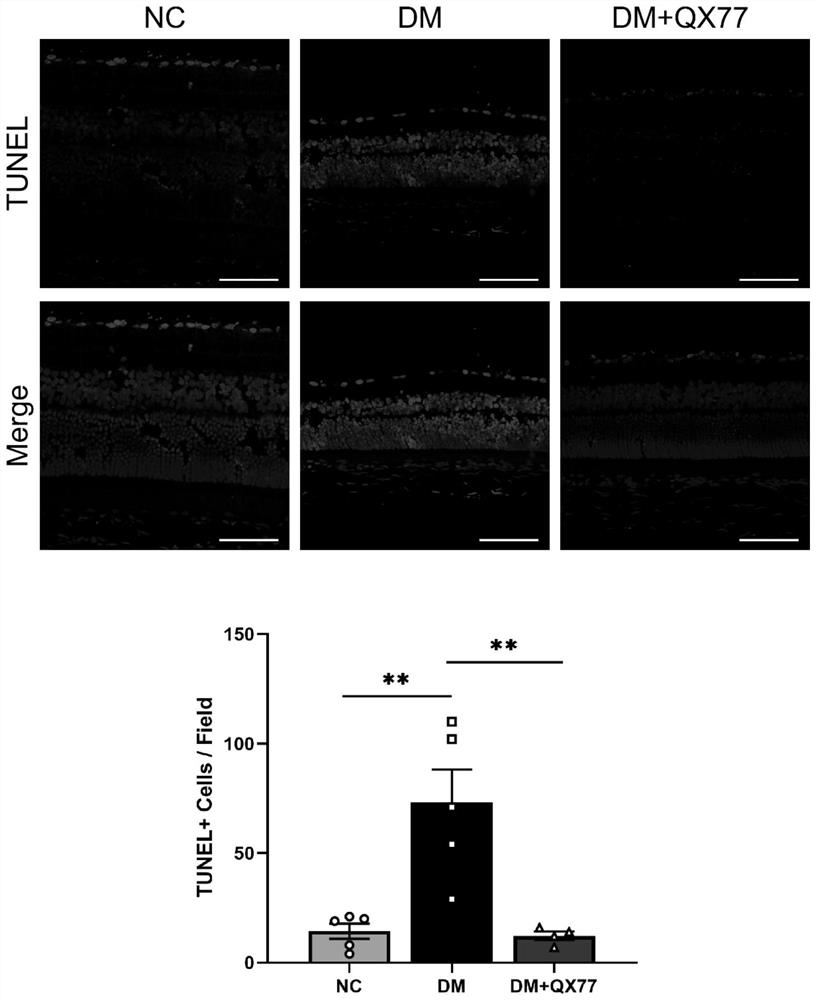
[0037] Example 3: Preparation of retinal frozen sections
[0038] 1.1 Eyeball preparation: Eyeball samples were collected from SD rats treated with drugs at different time points. After the SD rats were killed by dislocation, the eyeballs were carefully removed, and the optic nerve was preserved;
[0039] 1.2 Fix: place in 4% paraformaldehyde to fix overnight;
[0040] 1.3 Dissection: dissect the eyeball along the upper edge of the corneoscleral limbus under a dissecting microscope, carefully remove the cornea, iris and lens to form a complete optic cup, and pay attention to gentle operation to prevent retinal detachment;
[0041] 1.4 Dehydration: overnight dehydration with 30% sucrose;
[0042] 1.5 Embedding: embed with tissue embedding solution OCT at 4°C and equilibrate overnight;
[0043] 1.6 Quick freezing with liquid nitrogen: Quickly freeze the eyeballs with liquid nitrogen, and make the eyeballs as vertically centered as possible before freezing. Store frozen sample...
PUM
 Abstract
Description
Claims
Application Information
 - R&D
- Intellectual Property
- Life Sciences
- Materials
- Tech Scout
- Unparalleled Data Quality
- Higher Quality Content
- 60% Fewer Hallucinations
Browse by: Latest US Patents, China's latest patents, Technical Efficacy Thesaurus, Application Domain, Technology Topic, Popular Technical Reports.
© 2025 PatSnap. All rights reserved.Legal|Privacy policy|Modern Slavery Act Transparency Statement|Sitemap|About US| Contact US: help@patsnap.com