
Preparation method of electrically functionalized graphene electron microscope grid

A graphene film, single-layer graphene technology, applied in chemical instruments and methods, circuits, discharge tubes, etc., can solve the problem of single orientation of the sample on the support film, and achieve the effect of convenient acquisition, simple method, light and thin conductivity

Pending Publication Date: 2022-06-24
SHUIMU BIOSCIENCES LTD +1
View PDF0 Cites 0 Cited by
  • Summary
  • Abstract
  • Description
  • Claims
  • Application Information

AI Technical Summary

Problems solved by technology

As an ultra-thin two-dimensional material, graphene film combines the advantages of porous carbon film and continuous carbon film by virtue of its high transparency, high conductivity and good mechanical strength, which can eliminate the problems existing in the current electron microscope grid to varying degrees. Strong background noise, poor conductivity, distribution of biomolecules at the gas-liquid interface, and sample drift under electron beam irradiation, but the problem of single orientation of the sample on the support film is difficult to solve, and this problem has become a constraint for single particle analysis of high-resolution structures. Parsing the main bottleneck

Method used


Image

  • Preparation method of electrically functionalized graphene electron microscope grid
  • Preparation method of electrically functionalized graphene electron microscope grid
  • Preparation method of electrically functionalized graphene electron microscope grid

Examples


Embodiment 1

[0100] Embodiment 1, the preparation of electrically functionalized graphene electron microscope carrier network

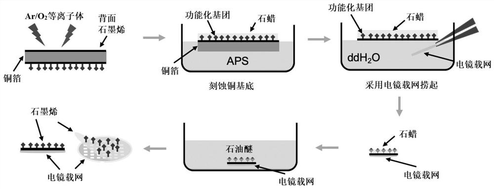
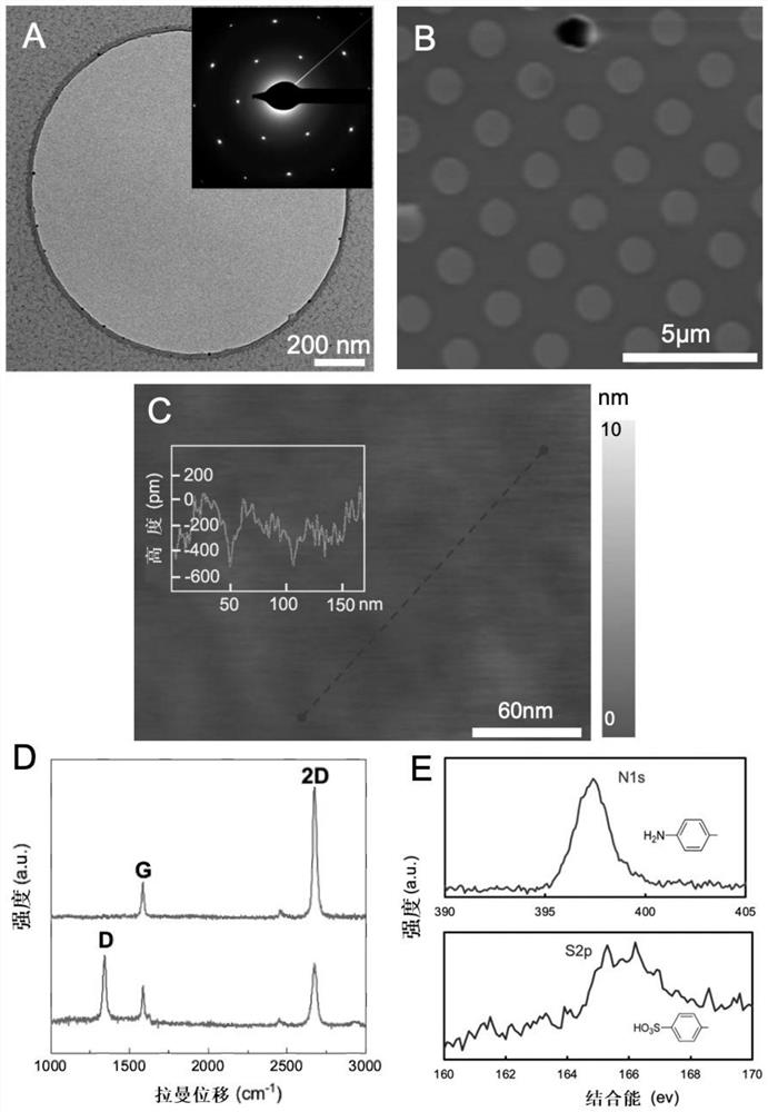
[0101] figure 1This is a schematic diagram of the preparation of electrically functionalized graphene electron microscope carrier network. The principle is to use the reaction between diazonium salt and graphene carbon skeleton to first covalently modify the graphene grown on the copper foil, so that the graphene film can be in the buffer solution. The positive and negative charges are respectively ionized, and then the graphene monolayer film is transferred to the electron microscope carrier grid with paraffin, and the graphene electron microscope carrier grid can be obtained after the paraffin is removed with an organic solvent. The specific process is as follows:

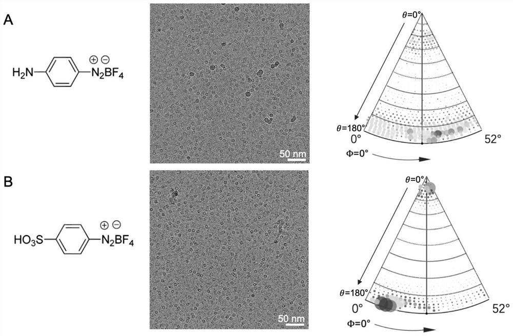
[0102] 1. Synthesis of diazonium salts with different electrical functional groups

[0103] Synthesize 4-aminobenzene tetrafluoroborate diazonium salt and 4-sulfonic acid benzene tetrafluorobora...

Embodiment 2

[0112] Embodiment 2, the application of electrically functionalized graphene electron microscope carrier network

[0113] Take the 20S proteasome, the ribosome and the second intron LtrB RNP as examples to make frozen samples.

[0114] First, 5 μL of protein buffer (25 mM Tris-HCl, 150 mM NaCl, pH 8.0) was added dropwise to the amino-functionalized and sulfonic acid-functionalized graphene electron microscope carriers prepared in Example 1, and incubated for 2 min to activate the amino groups on the graphene membrane. and sulfonate ions, then the buffer was removed with filter paper, about 4 μL of 20S proteasome solution was added dropwise, and then transferred to FEIVitrobot (refrigerated sample preparation instrument) for frozen sample preparation. The humidity of the Vitrobot cavity was adjusted to 100%, the temperature was adjusted to 12°C, the suction time of the filter paper was set to 1.5s-2s, and the force was set to -2. After removing the excess liquid with filter ...


PUM

PropertyMeasurementUnit
melting pointaaaaaaaaaa
densityaaaaaaaaaa

Abstract

The invention discloses a preparation method of a different-electrical-property functionalized graphene electron microscope grid, and relates to a method for functionalizing a graphene film by using functional groups with different electrical properties and transferring the functionalized graphene film to an electron microscope grid. The method comprises the following steps: reacting a graphene film with diazonium salt, paving a hydrocarbon mixture, transferring the graphene film to an electron microscope grid, and removing the hydrocarbon mixture. The functionalized graphene electron microscope grid prepared by the invention can keep the advantage of lower background noise of graphene as an electron microscope grid support film, the support film has an enrichment effect on samples, biomacromolecules are not distributed to a gas-liquid interface to deform, and meanwhile, the problem of dominant orientation can be solved. The graphene film with different electrical functionalizations, provided by the invention, can bring great convenience to sample preparation and later three-dimensional reconstruction of a cryoelectron microscope.

Description

technical field [0001] The invention relates to a method for preparing a functionalized graphene electron microscope carrier network with different electrical properties. A single-layer graphene film is modified with groups of different charges, and an electron microscope carrier network covered by a modified graphene supporting film is prepared to change the properties of protein particles. The problem of dominance orientation belongs to the technical field of biological cryo-electron microscopy. Background technique [0002] Since the development of cryo-electron microscopy, it has become an indispensable means of structural biology research. Single particle analysis is currently the mainstream method for structural analysis of biological macromolecules using cryo-electron microscopy. Single particle analysis is based on the assumption of molecular structure identity, and uses the random distribution of biomolecules to perform statistical analysis and reconstruction of mu...

Claims


Application Information

Patent Timeline
Patent Type & Authority Applications(China)
IPC IPC(8): H01J37/20H01J37/26C01B32/194
CPCH01J37/20H01J37/26C01B32/194
Inventor 王宏伟郭春龙饶燏刘楠陆叶刘永波倪晓丹
Owner SHUIMU BIOSCIENCES LTD
Learn More