Assay and kit for detection of endotoxin

a technology of endotoxin and kit, which is applied in the field of assays, devices and kits for detection of endotoxin in samples, can solve the problems of endothelial cell destruction, inflammatory reactions and septic shock in patients, and the growth of contaminant bacteria that produce toxins, etc., and achieve the effect of rapid detection of endotoxins

Active Publication Date: 2021-04-06
NANODX HEALTHCARE PVT LTD
View PDF4 Cites 0 Cited by
  • Summary
  • Abstract
  • Description
  • Claims
  • Application Information

AI Technical Summary

Benefits of technology

[0009]Accordingly, the present invention in general aspects provides a membrane based assay method, device and kit for rapid detection of endotoxins in a sample.
[0014]In an embodiment, the membrane based assay method for detection of endotoxins as per the present invention optionally comprises a step of adding a protein solution following the placement of the sample over the same area for minimizing any non-specific binding in the subsequent steps.
[0016]In one embodiment, the membrane based assay method for detection of endotoxins as per the present invention optionally comprises adding ultrapure water free of endotoxins over the coloured signal appeared subsequent to the placement of the suspension of conjugates of LPS-affinity ligand with GNPs for removing any excess unbound GNPs and improving the accuracy of determination of endotoxin levels in the sample.

Problems solved by technology

Exposure to endotoxins associated with Gram-negative bacteria (GNB) can lead to effects such as fever, cytokine system activation, endothelial cell destruction, blood vessel permeability, or other adverse effects.
All parenteral and biological pharmaceutical products and processed food items require rapid pyrogen testing for safety an quality assurance as otherwise, the presence of endotoxin in injectable and oral formulations and food products can lead to inflammatory reactions and septic shock in patients.
Moreover, improper storage conditions of food items can lead to growth of contaminant bacteria that produce toxins.
Moreover, sepsis is the single largest cause of hospital-acquired infections and fatalities especially in resource-poor settings.
Despite advances in antimicrobial therapy, septic shock and other clinical complications due to GNB infections continue to pose a major health problem.
The delay in initiation and / or ineffectiveness of appropriate antibiotic therapies can lead to fatalities.
Commonly applied sepsis detection methods based on microbiological analysis are hampered by the uncertainties associated with the growth of bacterial pathogens in culture media due to prior antibiotic exposure, while newer methods based on detection of procalcitonin (PCT) levels, a surrogate sepsis biomarker, in serum are unable to offer definitive results.
Initially for many years, bacterial endotoxin tests (BETs) were performed by the USP Rabbit Pyrogen Test, a time-consuming and costly animal test.
LAL test and its many versions are time consuming and need expensive equipments like incubators (heating and heat control unit), photometers, a special machine to detect the test results, e.g., a microplate reader, etc.
These methods need specialized technical expertise, are required to be performed inside a laboratory and require expensive equipments increasing the cost per test.

Method used


Image

  • Assay and kit for detection of endotoxin
  • Assay and kit for detection of endotoxin
  • Assay and kit for detection of endotoxin

Examples


example

Example 1

[0111]Membrane Device:

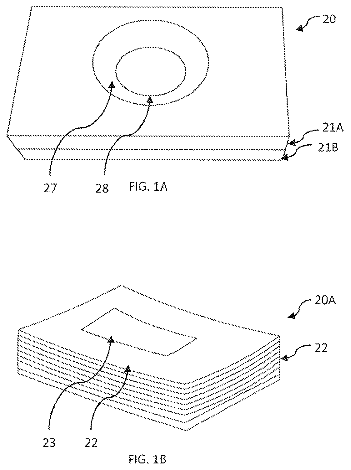
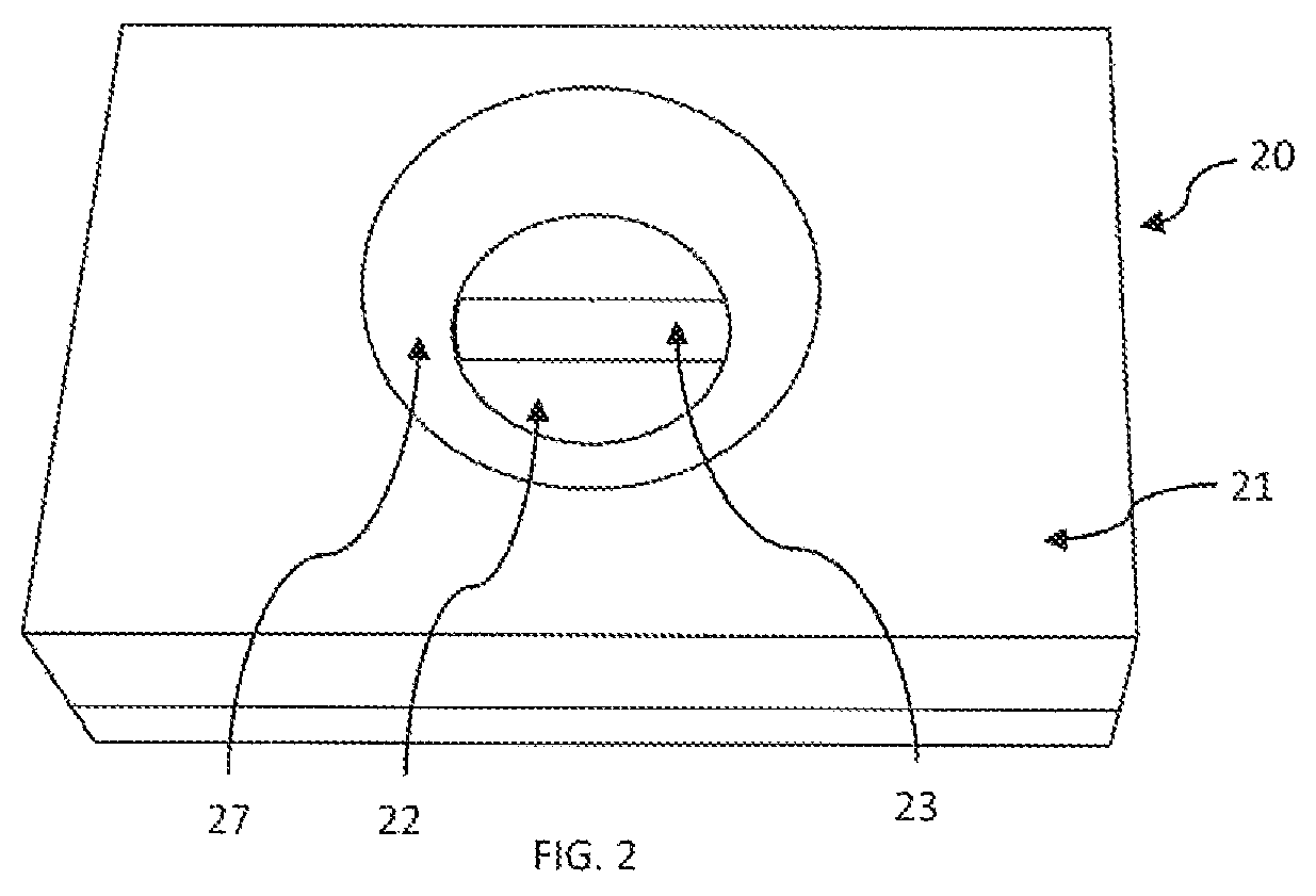
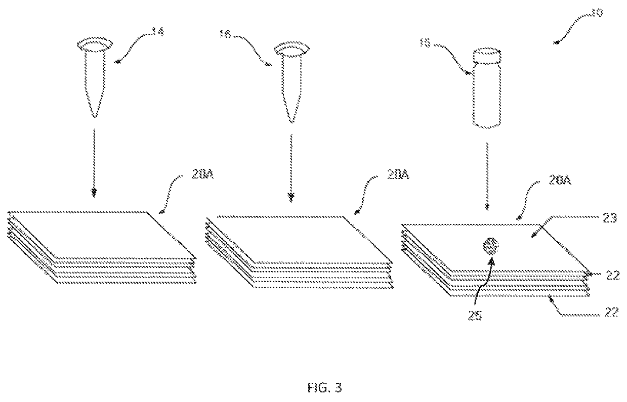
[0112]Plastic cassette 21 (FIG. 1A) of 4 cm×3 cm×1 cm dimension made by injection-molding having an upper component 21A and lower component 21B and was used as an enclosure for enclosing membranes therein. As shown in FIG. 2, in the lower component 21B multiple layers of membranes 20A as shown in FIG. 1B were placed. The multiple layers of membranes 20A included a stack of high wicking capacity absorbent pads 22 type-AP080 (Advanced Microdevice Pvt. Ltd. Amabala, India), over which was placed an endotoxin-affinity membrane 23 of nitrocellulose type-SCNM; pore size 0.22 μm; (Advanced microdevice Pvt. Ltd., Amabala, India). The upper component 21A is provided with an opening 28 created by the concave depression 27 which firmly holds the membranes in the component 21B. The opening 28 allows placing of the sample and other reagents through it over the endotoxin-affinity membrane 23. The membrane device 20 when constructed appeared as seen in FIG. 2.

example 2

[0113]Synthesis of Citrate-Stabilized GNPs:

[0114]Citrate-stabilized GNPs of size 16±4 nm were synthesized by following the method as reported in the literature mentioned hereinabove in the detailed description.

[0115]Briefly, HAuCl4.3H2O was reduced with an aqueous solution of sodium citrate and tannic acid. 10 mL of 1% (w / v) HAuCl4 solution was diluted to 800 ml with uitrapure DI water (solution A) and another aqueous solution containing 40 mL of 1% (w / v) sodium citrate and 100 μL of 0.1% (w / v) tannic acid was diluted to 200 mL (solution B), Both A and B solutions were mixed at 60° C. and kept for 4 h under constant stirring. The mixture changed its colour from light yellowish to black to violet and finally to red. The mixture was removed from heat and immediately chilled in an ice bath to quench the reaction. The prepared AuNP suspension was then stored at 4° C. until further use. The stability of the AuNP suspension was checked for 3 consecutive months by UV-Visible spectroscopy a...

example 3

[0116]Preparation of Polymyxin B (PMB) Sulfate Antibiotic Drug Conjugated GNPs:

[0117]Polymyxin B (PMB) sulfate antibiotic drug conjugated GNPs were prepared by the protocol as reported by Kalita et al as follows:

[0118]The covalent conjugation of PMB to GNPs was performed in three steps. 10 mL of 18 nM aqueous GNPs suspension was incubated at room temperature with 100 μL, of 18 mM DTH in ethanol for 8 h under constant stirring at 50 rpm using rotospin. This allowed the DTH to ligand place exchange with the surface-capping citrate groups on gold. Excess DTH was removed by centrifuging three times at 5080, 7040 and 17540 rcf (g force), respectively, for 15 min each and the particles were resuspended in 20 mL of 2.5% v / v GLA in HEPES buffer. The suspension was left for incubation at room temperature overnight under constant stirring at 50 rpm using rotospin. The amine reactive conjugates obtained at this stage were washed thrice by centrifugation as described above and the NP pellet was...


PUM

PropertyMeasurementUnit
concentrationaaaaaaaaaa
diameteraaaaaaaaaa
thicknessaaaaaaaaaa

Abstract

The present invention relates to a membrane based assay method, device and kit for rapid detection and / or quantification of endotoxins in aqueous solutions and test samples. The kit as per the present invention comprises lipopolysaccharide (LPS) affinity ligand conjugated with gold nanoparticles (GNPs); a membrane device comprising an endotoxin affinity membrane positioned parallelly to one or more layer(s) of a hydrophilic material, which are optionally secured in an enclosure; and optionally comprising an indicator chart for quantification of endotoxins in the sample. The method comprises placing the sample suspected of endotoxin contamination on a surface of a membrane comprised in a membrane device; placing once or more a suspension of LPS-affinity ligand conjugated with GNPs over the same area as the sample placed and detecting the presence of endotoxin if the colour signal appears and based on its intensity quantifying the endotoxin levels.

Description

FIELD OF THE INVENTION[0001]The present invention relates to an assay, a device and a kit for detection of endotoxin in a sample. More particularly, the present invention relates to a membrane based assay method, device and kit for rapid endotoxin detection in a sample.BACKGROUND OF THE INVENTION[0002]Endotoxin detection in test samples from pharmaceutical and food industries as well as in clinical and healthcare setup is of paramount importance. Exposure to endotoxins associated with Gram-negative bacteria (GNB) can lead to effects such as fever, cytokine system activation, endothelial cell destruction, blood vessel permeability, or other adverse effects. Endotoxin or lipopolysaccharide (LPS) present on the cell wall of GNB is responsible for many toxic effects that occur during GNB sepsis and plays an important role in the pathophysiology of such infections.[0003]All parenteral and biological pharmaceutical products and processed food items require rapid pyrogen testing for safety...

Claims


Application Information

Patent Timeline
Patent Type & Authority Patents(United States)
IPC IPC(8): G01N33/569G01N33/543G01N33/531G01N33/58
CPCG01N33/56911G01N33/531G01N33/543G01N33/54366G01N33/587G01N2400/50G01N33/54386G01N33/54388
Inventor GUPTA, SHALINISRITHARAN, VENKATARAMANKALITA, PRASANTA
Owner NANODX HEALTHCARE PVT LTD
Why Eureka
Try Eureka