Detection of micro metastasis of melanoma and breast cancer in paraffin-embedded tumor draining lymph nodes by multimarker quantitative RT-PCR

a paraffin-embedded tumor and micro-metastasis technology, applied in the field of paraffin-embedded tumor micro-metastasis detection, can solve the problems of malignant neoplasms' tendency to disseminate cells, high labor intensity of approaches, and often underestimate the presence of micro-metastatic disease in procedures. , to achieve the effect of accurate determination, high sensitivity and specificity

Inactive Publication Date: 2004-12-30
JOHN WAYNE CANCER INST
View PDF10 Cites 11 Cited by
  • Summary
  • Abstract
  • Description
  • Claims
  • Application Information

AI Technical Summary

Benefits of technology

0021] The present invention provides a number of unexpected advantages. First, it allows to determine stages of the cancers more accurately as compared to the existing methods. The methods and kit of the present invention allow accurate determination of a need for further lymph node removal surgery and/or adjuvant treatment. Second, the present invention demonstrates the better reliability of molecular marker panels of the present inve

Problems solved by technology

One of the most devastating aspects of cancer is the propensity of cells from malignant neoplasms to disseminate from their primary site to distant organs and develop into metastases.
However, such approach is labor-intensive.
Also, about 80% of patients who undergo elective lymph node dissection have no evidence of metastases and do not benefit from this procedure.
But this procedure often underestimates the presence of micrometastatic disease.
Despite initially encouraging results, the clinical and survival significance of these occult metastases detected by RT-PCR assays has not been demonstrated conclusively.
Furthermore, conventional single mRNA marker RT-PCR assays are limited in their ability to discriminate cancer cells from normal cells that also carry the marker and, thus, these methods suffer from reduced specificity and reliability.
In addition, tumor heterogeneity has caused sensitivity problems where a single mRNA marker was employed--although all tumor cells within a primary tumor or metastasis may express the same marker gene, the level of specific mRNA expression can vary considerably.
Thus, despite the identification of melanoma and breast cancer markers, these markers cannot individually detect tumor cells in a highly specific and sensitive manner (U.S.

Method used


Image

  • Detection of micro metastasis of melanoma and breast cancer in paraffin-embedded tumor draining lymph nodes by multimarker quantitative RT-PCR
  • Detection of micro metastasis of melanoma and breast cancer in paraffin-embedded tumor draining lymph nodes by multimarker quantitative RT-PCR
  • Detection of micro metastasis of melanoma and breast cancer in paraffin-embedded tumor draining lymph nodes by multimarker quantitative RT-PCR

Examples


example 2

[0064] Histopathologic Examination and RNA Isolation

[0065] Histopathologic examination by the pathologists was performed on each collected SN as previously described (Bostick, P. J., 1999, which is incorporated herin by the reference). Each SN was bisected, and an 8-.mu.m imprint slide was then prepared from the tissue surface and stained with Diff-Quik I & II (Dade International, Miami, Fla.) for the pathologist's intraoperative diagnosis. If melanoma cells were identified in the frozen section, a complete lymph node dissection was then performed (Bostick, P. J., 1999). Six immediately adjacent frozen sections were cut on the cryostat to a thickness of 12 .mu.m each and stored at -80.degree. C. until processed at a later date (Bostick, P. J., 1999; Miyashiro, I., 2001). The remainder of the bisected node, which was not frozen, was then placed in 10% formalin and embedded in paraffin, and 4-.mu.m-thick sections were examined with H&E staining. Adjacent 4-.mu.m-thick sections werre e...

example 3

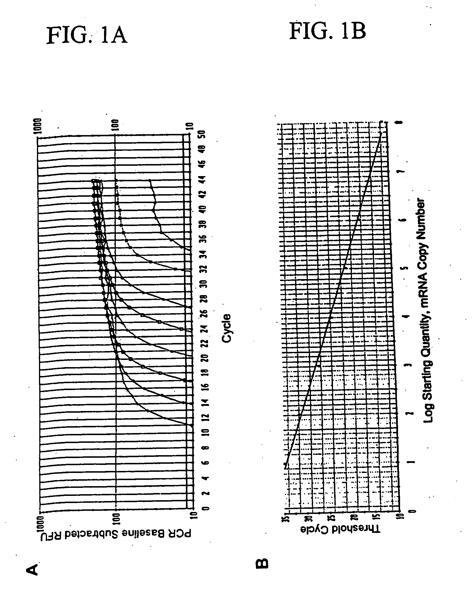
[0067] Dilution Study To verify the specificity of detecting occult metastasis, total RNA was extracted from 1000 melanoma cells of obviously metastatic melanoma tissue (n=8) in PE sections using laser capture microdissection (LCM). Five-.mu.m thick sections were cut from each tissue specimen embedded in paraffin using a microtomb and mounted on a slide. Then, tissue sections were deparaffinized with xylene and subsequently stained with H&E. After drying a slide, 1000 metastatic melanoma cells were microdissected using the PixCell II LCM System (Arcturus Engineering, Mountain View, Calif.) as manufacture's procedure. Microdissected tissues were digested with proteinase K and total RNA was extracted using the Paraffin Block RNA Isolation Kit (Ambion). RNA isolated from 1000 melanoma cells was dissolved in molecular grade water, and was serially diluted by 1 / 2, {fraction (1 / 10)}, {fraction (1 / 20)}, {fraction (1 / 50)}, {fraction (1 / 100)}, {fraction (1 / 500)}, and {fraction (1 / 1000)}. Rev...

example 4

[0068] Primers and Probes

[0069] Primer and probe sequences were designed for the RealTime PCR assay using Oligo Primer Analysis Software, version 5.0 (National Biomedical systems, Plymouth, Minn.). To avoid possible amplification of contaminating genomic DNA, primers were designed so that each PCR product covered at least one intron. Fluorescence Resonance Energy Transfer (FRET) probe sequences were as follows:

1 MART-1: 5'-FAM-CAG AAC AGT CAC CAC CAC CTT ATT-BHQ-1-3'; (SEQ ID NO:1) MAGE-A3: 5'-FAM-AGC TCC TGC CCA CAC TCC CGC CTG T-BHQ-1-3'; (SEQ ID NO:2) GalNAcT: 5'-CAL RED-ATG AGG CTG CTT TCA CTA TCC GCA-BHQ-2-3'; (SEQ ID NO:3) PAX3, 5'-FAM-CCA GAC TGA TTA CGC GCT CTC CC-BHQ-1-3'; (SEQ ID NO:4) Glyceraldehydes-3-phosphate dehydrogenase (GAPDH): 5'-FAM-CAG CAA TGC CTC CTG CAC CAC CAA-BHQ-1-3'. (SEQ ID NO:5)

[0070] Control melanomas and non-melanoma tissues and cell lines were used to optimize the assay. GAPDH was utilized as internal reference house-keeping genes for status of sample...


PUM

PropertyMeasurementUnit
Fractionaaaaaaaaaa
Fractionaaaaaaaaaa

Abstract

The invention provides a quantitative realtime RT-PCR assay for detection of metastatic breast, gastric, pancreas or colon cancer cells or metastatic melanoma. The assay allows to predict disease recurrence and survival in patients with AJCC stage I and II, and III disease using multimarker panels. The method for detecting metastatic melanoma cells utilizes panels of markers selected from a group consisting of MAGE-A3, GalNAcT, MART-1, PAX3, Mitf, TRP-2, and Tyrosinase. The method for detecting metastatic breast, gastric, pancreas or colon cancer cells in paraffin-embedded samples utilizes panels of markers selected from a group consisting of C-Met, MAGE-A3, Stanniocalcin-1, mammoglobin, HSP27, GalNAcT, CK20, and beta-HCG.

Description

[0001] This invention claims priority to the U.S. Provisional Patent Application No. 60 / 426,216 filed Nov. 14, 2002.[0002] The present invention relates to methods of detection of micro metastasis in lymph nodes and methods of predicting the recurrence of the disease and survival. More specifically, the invention is directed to detection of micro metastasis in cancerous tissue and lymph nodes by using multimarker real-time reverse transcriptase polymerase chain reaction assay.DESCRIPTION OF THE PRIOR ART[0003] The incidence of malignant melanoma has been increasing in the United States over the past decade (Balch, C. M., 2001; Jemal, A. J., 2002; and Rigel, D. S., 1996). Breast cancer is another common form of malignant disease whose incidence is increasing. One of the most devastating aspects of cancer is the propensity of cells from malignant neoplasms to disseminate from their primary site to distant organs and develop into metastases. Despite advances in surgical treatment of pr...

Claims


Application Information

Patent Timeline
IPC IPC(8): C12P19/34C12Q1/68G01N33/53
CPCC12Q1/6886C12Q2600/112C12Q2600/118
Inventor HOON, DAVE S.B.TAKEUCHI, HIROYA
Owner JOHN WAYNE CANCER INST
Why Eureka
Try Eureka