
Methods for detecting an analyte of interest using catalyzed reporter deposition of tyramide

a reporter and catalyzed technology, applied in the field of catalyzed reporter deposition of tyramide, can solve the problems of high level of spontaneous activation and non-specific binding of fluorescent substrates to live cell membranes, difficult direct detection of fluorochrome-labeled tyramide, etc., to achieve enhanced detection signal and improve catalyzed reporter deposition staining

Inactive Publication Date: 2005-01-06
VERVE
View PDF3 Cites 14 Cited by
  • Summary
  • Abstract
  • Description
  • Claims
  • Application Information

AI Technical Summary

Benefits of technology

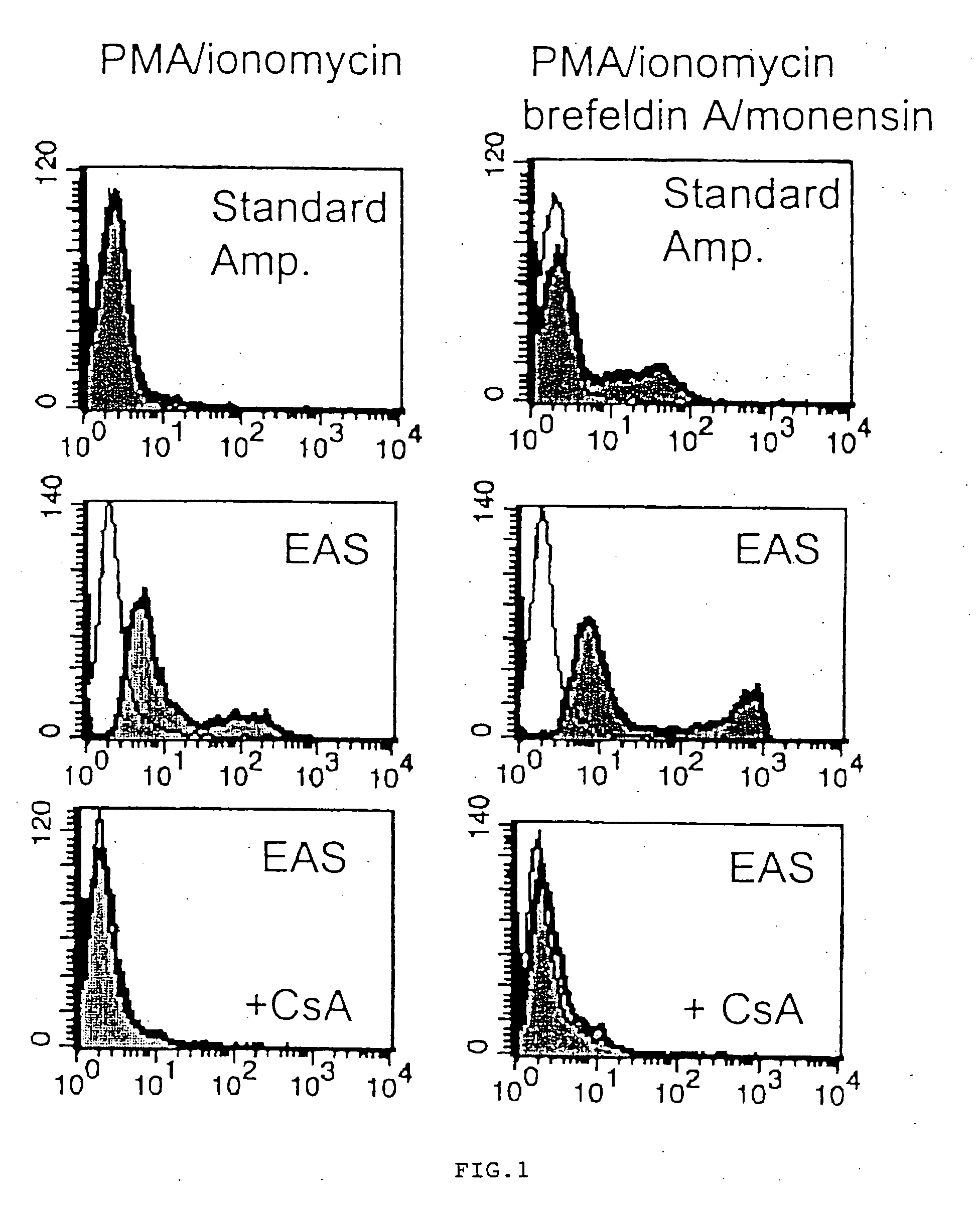
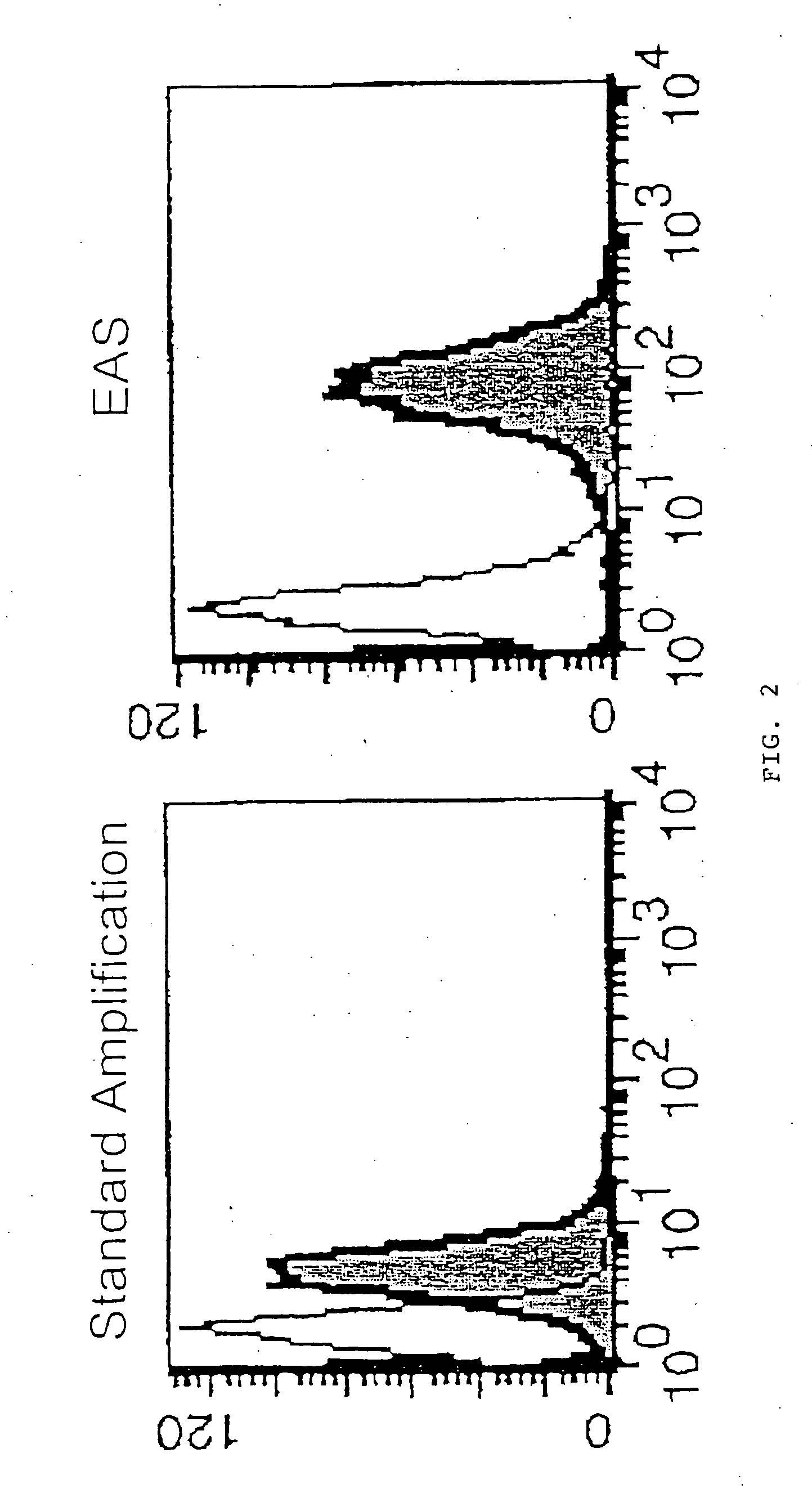
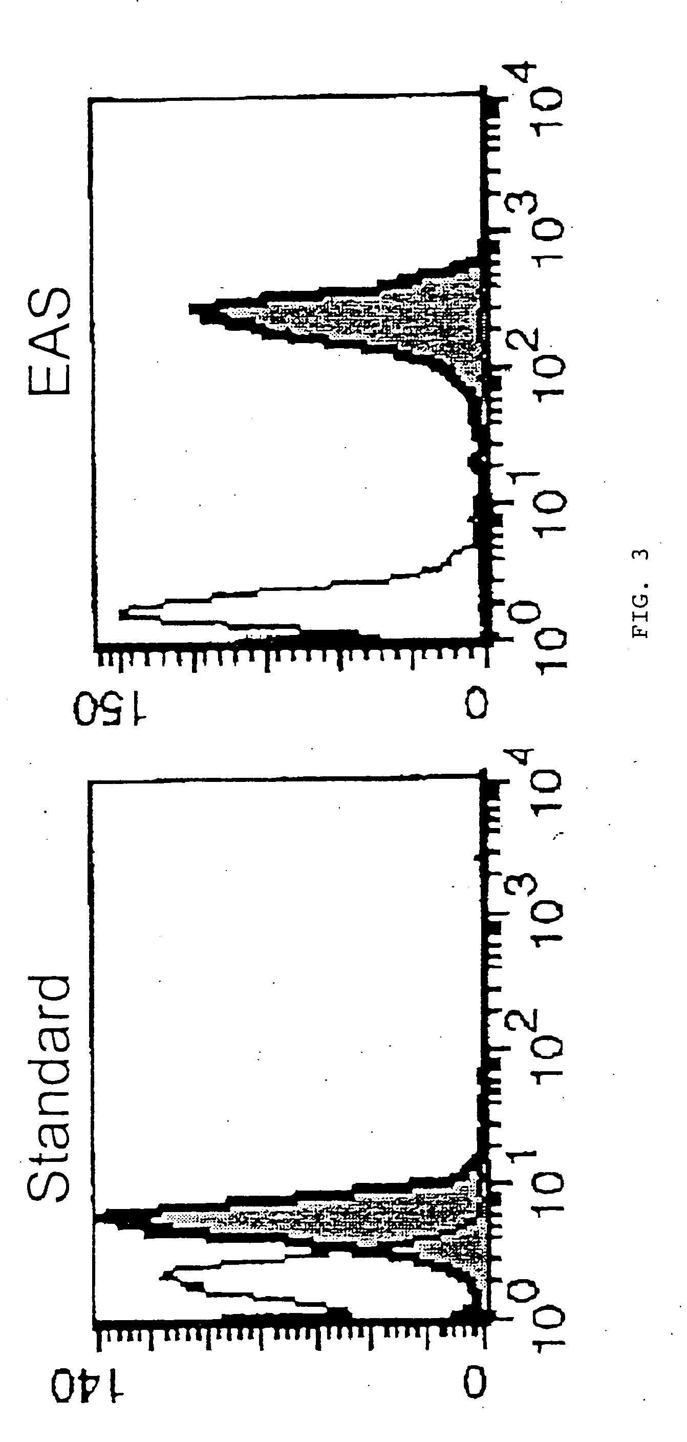
[0013] The invention provides methods for catalyzed reporter deposition staining of intracellular antigens that are simple, and that provide signal amplification levels of 10-fold or more in comparison to standard flow cytometry methods. Moreover, the invention also provides a method for tyramide coating cells for flow cytometry, wherein cells are preferably exposed to a catalyzed reporter deposition system which results in specific tyramide coating of cells which contain or express an analyte of interest. Additionally, the invention further provides materials and methods for improving catalyzed reporter deposition staining of antigens generally, including the staining of fixed sections, fixed and permeabilized cells, particles, and live cells.
[0025] By “aided existence” is meant adding components to the buffer or medium containing the cells which allows the cell to remain viable.
[0070] In yet another aspect, the present invention features compositions for use in catalyzed deposition methods. The compositions can provide reduced nonspecific backgrounds and greater peak separation between histograms obtained from cells stained with control immunoglobulin versus cells stained with immunoglobulin specific for an analyte of interest when used in flow cytometry, as well as in histochemical methods for staining analytes of interest present in thin sections, cell preparations, and other applications known to those of ordinary skill in the art.
[0076] In yet another aspect, the present invention features methods for optimizing compositions for use in catalyzed deposition methods by providing a library of amino acid, dipeptide and / or oligopeptide buffering agents as defined herein. In these methods, a plurality of buffering agents are compared for their ability to reduce nonspecific background staining and provide greater peak separation between histograms obtained from cells stained with control immunoglobulin versus cells stained with immunoglobulin specific for an analyte of interest when used in catalyzed reporter deposition methods. The methods can provide optimal compositions for use in flow cytometry, and in histochemical methods for staining analytes of interest present in thin sections, cell preparations, particles, and other applications known to those of ordinary skill in the art.

Problems solved by technology

Lollini et al. indicates that TSA is not superior to conventional techniques for detecting surface antigens on live cells, stating for example on page 5, “(t)he main problem appeared to be a high level of spontaneous activation and non-specific binding of the fluorescent substrate to live cell membranes.
The Karkmann et al. reference states that direct detection of fluorochrome-labeled tyramide is problematic, “probably due to hydrophobic interactions with the cell membrane” Thus, the Karkmann et al. reference uses a complicated indirect staining method, in which antibody selective for an antigen of interest, coupled to peroxidase, catalyzes the deposition of biotin-tyramide, which is detected by fluorescently labeled streptavidin.

Method used


Image

  • Methods for detecting an analyte of interest using catalyzed reporter deposition of tyramide
  • Methods for detecting an analyte of interest using catalyzed reporter deposition of tyramide
  • Methods for detecting an analyte of interest using catalyzed reporter deposition of tyramide

Examples


example 1

Detection of Cytokines

[0115] Human peripheral blood mononuclear cells were cultured in RPMI 1640 medium +10% fetal bovine serum, containing 10 ng / ml phorbol myristic acetate and 500 ng / ml ionomycin, with or without 3 μM monensin and 10 μM brefeldin A, at 37° C. for 5 hours to induce expression of Interleukin 2. In some cultures, 10 μg / mL of cyclosporin A (an inhibitor if IL-2 induction) was included.

[0116] The cells were then harvested and fixed in 100 μL of 1% paraformaldehyde in phosphate buffered saline (PBS) at room temperature for 10 minutes, then permeabilized overnight at room temperature in 100 μL of 0.2% saponin, 3% hydrogen peroxide, and 1 mg / mL milk proteins in PBS. The cells were then washed in a PBS diluent (consisting of PBS with 1% bovine serum albumin and 1% fetal bovine serum).

[0117] The cells were then treated for 10 minutes at room temperature with 500 ng mouse IgG1 or 500 ng mouse IgGI1 anti-human interleukin 2 primary antibody (R&D Systems).

[0118] After bein...

example 2

Detection of Intracellular Antigens

[0123] Approximately one million cells were washed in PBS, followed by incubation with 0.1 mL of 1% paraformaldehyde in PBS for 10 minutes at room temperature to fix the cells.

[0124] The cells were washed in PBS and then incubated with 0.1 ml of 0.2% saponin, 3% hydrogen peroxide, and 1 mg / mL milk proteins in PBS for 10 minutes at room temperature. This step permeabilizes the cells, inhibits endogenous peroxidases, and blocks nonspecific binding.

[0125] The cells were washed in a PBS diluent (consisting of PBS with 1% bovine serum albumin, 1% fetal bovine serum, which may or may not contain 0.2% saponin). The cells were then incubated for 10 minutes at room temperature with 0.05 mL of an unconjugated primary antibody in the same PBS diluent. The concentration of the antibody can vary, depending on the antibody used. Optimal antibody concentration is determined empirically. Unbound antibody is then washed out of the cells with the PBS diluent.

[01...

example 3

Detection of Cellular RNA Sequences

[0130] Approximately one million cells were incubated for 10 min at about 22° C. in 100 μL of a solution consisting of 2% paraformaldehyde in phosphate-buffered saline (PBS). The cells were washed once in PBS, followed by a 15 minute incubation at about 22° C. in 1 ml of a solution consisting of 0.2% diethyl-pyrocarbonate in 50% ethanol / 50% PBS. This solution was made fresh daily. For all subsequent steps, all solutions were treated with 0.2% diethyl-pyrocarbonate for 10 min. at about 22° C. to destroy any RNase present, and then autoclaved to destroy the chemical.

[0131] The cells were subsequently washed twice with PBS and then incubated for 10 min at about 22° C. in 3% hydrogen peroxide and 1 mg / ml milk proteins in PBS. After this step, the cells were washed in 4×SCC containing 5 mM sodium phosphate, pH 7.4, and 50% formamide. The 4×SSC was diluted from a 20×SCC stock (175.3 grams sodium chloride +88.2 grams sodium citrate +00 ml deionized (DEP...


PUM


Abstract

The present invention relates to methods of staining cells for flow cytometry, using catalyzed reporter deposition and amplification staining. A catalyzed reporter deposition or an analyte dependent enzyme activation system is described for detecting and / or quantitating an analyte of interest in a cell by flow cytometry. Also described are compositions for use in catalyzed reporter deposition methods that can be used to reduce background staining, and thereby enhance peak signal separation between histograms obtained from cells stained with control immunoglobulin versus cells stained with immunoglobulin specific for an analyte of interest, in catalyzed deposition methods.

Description

[0001] This application is a continuation of U.S. application Ser. No. 09 / 835,287, filed on Apr. 13, 2001, which is a continuation-in-part of U.S. application Ser. No. 09 / 738,049, filed on Dec. 15, 2000, which are hereby incorporated by reference in their entireties, including all figures and claims.INTRODUCTION [0002] The present invention relates in part to methods of using tyramide in order to enhance the detection of analytes by flow cytometry, preferably using catalyzed reporter deposition and amplification staining. In preferred embodiments, the analytes detected according to the instant methods are cellular antigens, which may be intracellular or expressed on the surface of cells. The invention also relates in part to methods and compositions for use in amplification staining of analytes. BACKGROUND OF THE INVENTION [0003] The following discussion of the background of the invention is merely provided to aid the reader in understanding the invention and is not admitted to desc...

Claims


Application Information

Patent Timeline
IPC IPC(8): G01N1/30G01N33/569
CPCG01N2001/302G01N33/56972
Inventor KAPLAN, DAVID R.
Owner VERVE
Learn More