Compounds or agents that inhibit and induce the formation of focal microvessel dilatations

a microvessel and focal microvessel technology, applied in the field of inflammation modulation, can solve the problems of no conception of the potential use of pro-angiogenic factors in cancer treatment, and achieve the effects of broadening the potential for development of therapeutics, promoting lymphocyte transmigration, and preventing pathologies involving lymphocytic inflammation

Inactive Publication Date: 2005-06-30
DANA FARBER CANCER INST INC
View PDF1 Cites 11 Cited by
  • Summary
  • Abstract
  • Description
  • Claims
  • Application Information

AI Technical Summary

Benefits of technology

[0007] The invention is based on the discovery that within the microcirculation an anatomical change occurs that promotes lymphocyte transmigration across the vascular endothelium. Herein, it is shown that lymphocyte slowing and transmigration in the skin are associated with focally dilated vascular segments termed “microangiectasias,” also referred to herein as “focal microvessel dilatations.” The formation of focal microvessel dilatations in the microvasculature broadens the potential for development of therapeutics to treat both cancer and inflammatory disorders. In the present invention, methods of screening for agents or compounds that inhibit or induce the formation of focal microvessel dilatations are described. In addition, methods for the treatment of cancer, as well as, methods for both the treatment and prevention of pathologies involving lymphocytic inflammation are disclosed.
[0009] Angiogenesis is the proliferation of new blood vessel growth whose progression occurs in several phases that include; the presence of an angiogenic signal, dissolution of the blood vessel basement membrane, endothelial cell proliferation, endothelial cell migration, and the formation and differentiation of capillary tubules and loops. Pro-angiogenic factors thus can be used to induce endothelial cell proliferation and potentiate focal microvessel dilatation formation thereby allowing for lymphocyte transmigration.
[0013] In one embodiment, the method further comprises the step of administering the compound to an animal and determining whether focal microvessel dilatation formation is inhibited at a site of inflammation. In another embodiment, the inhibition of focal microvessel dilatation reduces the accumulation of perivascular lymphocytes at the site of inflammation. In another embodiment, the method further comprises administering an inhibitor of lymphocyte cell-cell adhesion.
[0017] In one embodiment, the method further comprises the step of administering the compound to an animal with a tumor and detecting the formation of focal microvessel dilatations adjacent to the tumor. In another embodiment, the induction of the focal microvessel dilatations increases perivascular lymphocyte accumulation within the tumor.

Problems solved by technology

However, until now, there has been no conception for the potential use of pro-angiogenic factors in the treatment of cancer.

Method used


Image

  • Compounds or agents that inhibit and induce the formation of focal microvessel dilatations
  • Compounds or agents that inhibit and induce the formation of focal microvessel dilatations
  • Compounds or agents that inhibit and induce the formation of focal microvessel dilatations

Examples


example 1

Lymphocyte Slowing and Transmigration in Focal Microvessel Dilatations

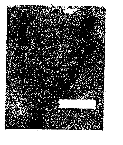
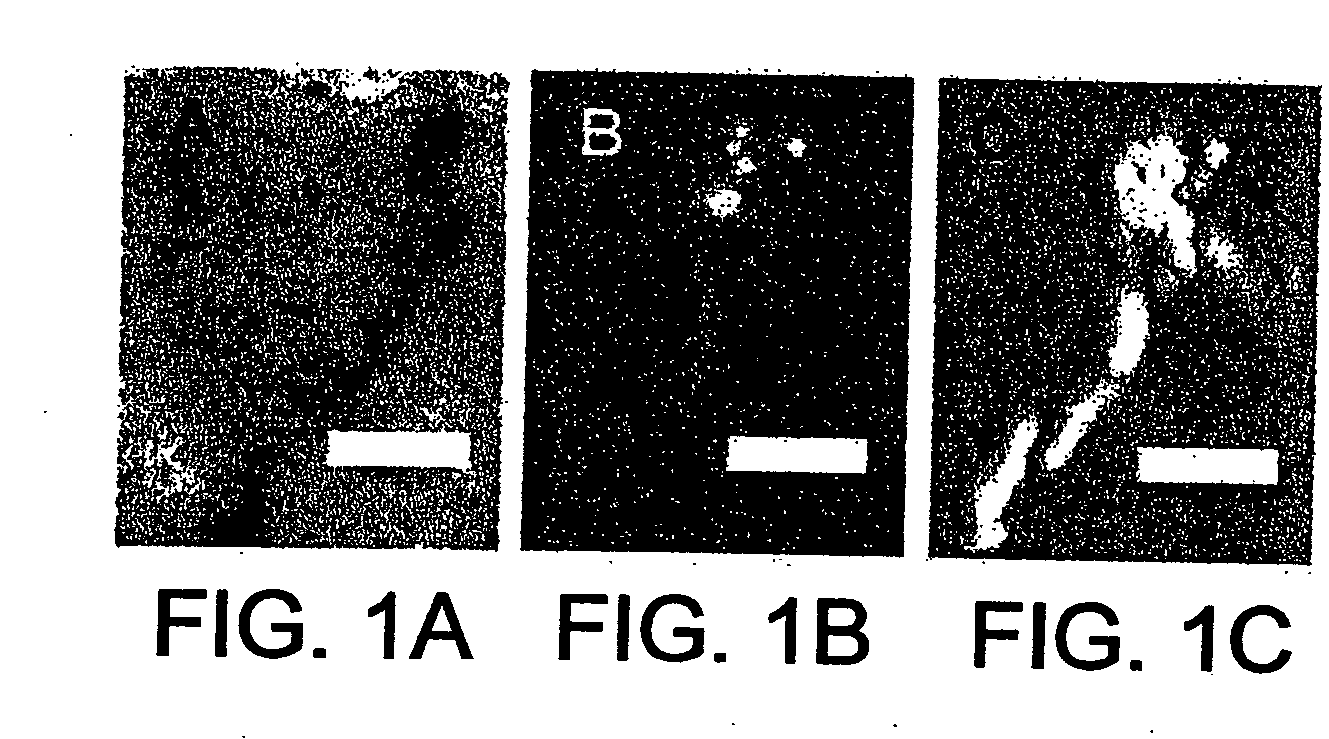
[0121] The intravital videomicroscopy studies demonstrated reproducible lymphocyte slowing in focal regions of the microcirculation (FIG. 1). Lymphocytes in these regions demonstrated a greater than 10-fold reduction in flow velocity (FIGS. 2 and 3). After the cells passed through these vascular segments, they rapidly returned to baseline flow velocities (FIG. 4). Also suggesting discrete structural changes in the sheep skin microcirculation, the regions of lymphocyte slowing were identified at approximately 100 um intervals (FIG. 1). The focal areas defined not only areas of lymphocyte slowing, but also the regions of lymphocyte transmigration. The focal areas of lymphocyte slowing were the only regions of the superficial vascular plexus where lymphocyte transmigration was observed (FIG. 1) (West et al. (2001), Am. J. Physiol. Heart Circ. 281, H1742-H1750. These findings suggested that lymphocyte transmigration ...

example 2

Focal Microvessel Dilatation Morphology

[0122] To evaluate the morphology of these focal regions of lymphocyte slowing and transmigration, corrosion cast injections of the inflammatory microcirculation were performed. The corrosion casts were examined by scanning and transmission electron microscopy and evaluated by digital morphometry. After systemic heparinization with 750 u / kg intravenous heparin, the external auricular arteries were bilaterally cannulated and perfused with approximately 100 cc of 37° C. saline followed by a 2.5 percent buffered glutaraldehyde solution (Sigma) at pH 7.40. The casts were made by perfusion of the ear arteries with 100 cc of Mercox (SPI, West Chester Pa.) diluted with 20 percent methylmethacrylate monomers (Aldrich Chemical, Milwaukee Wis.). After complete polymerization, the ears were harvested and macerated in 5% potassium hydroxide followed by drying and mounting for scanning electron microscopy. The microvascular corrosion casts were imaged afte...

example 3

Focal Microvessel Dilatation Wall Shear Stress and Microhemodynamic Mapping of Focal Microvessel Dilatations

[0124] The focal structural changes and the reduction in flow velocity indicated that focal microvessel dilatations have a significant impact on wall shear stress in the microcirculation. To define the microhemodynamic implications of the focal microvessel dilatations, the corrosion casts of the inflammatory skin were evaluated by quantitative 3-dimensional (3-D) scanning electron microscopy (Konerding, et al. (2001), Br J Cancer 84, 1354-62, M. A. Konerding et al. (1999), Br J Cancer 80, 724-32). Based on these data, 3-D hemodynamic maps of the focal microvessel dilatations were calculated. Wall shear stresses in the focal microvessel dilatations demonstrated a greater than 10-fold reduction in wall shear stress (FIG. 6). To estimate wall shear stresses, finite-element computations of flow fields in the neighborhood of the transition from the afferent vessel to the dilated s...


PUM


Abstract

The invention relates to the discovery that inflammation involves a structural modification to the microvascular architecture that results in the formation of focal microvessel dilatations or “microangiectasias.” The focal microvessel dilatations cause localized slowing of blood cell flow velocity that promotes the accumulation of extravascular lymphocytes. Methods are disclosed herein that exploit this discovery to screen for compounds that modulate the accumulation of extravascular lymphocytes. In addition, methods for treating patients suffering from cancer and diseases or pathologies associated with lymphocytic infiltration are disclosed.

Description

RELATED APPLICATIONS [0001] This application is a Continuation of PCT / US03 / 17790, filed Jun. 6, 2003, which claimed priority to U.S. Provisional Application No. 60 / 386,831, filed Jun. 6, 2002; the entirety of each of which is incorporated herein by reference.GOVERNMENT GRANTS [0002] At least part of the work contained in this application was performed under NIH grant HL47078. The government may have certain rights in this invention.FIELD OF THE INVENTION [0003] This invention relates to the modulation of inflammation. BACKGROUND OF THE INVENTION [0004] When recirculating lymphocytes migrate from the microcirculation to the extravascular site of inflammation, they must overcome the mechanical forces produced by blood flow. Blood flowing across the vascular endothelium creates shear forces at the endothelial boundary that are dependent on both flow velocity and vessel geometry. These shear forces disrupt the lymphocyte-endothelial cell adhesions necessary for transmigration. For more ...

Claims


Application Information

Patent Timeline
Patent Type & Authority Applications(United States)
IPC IPC(8): A61K49/00
CPCA61K49/0004
Inventor MENTZER, STEVEN J.
Owner DANA FARBER CANCER INST INC
Why Eureka
Try Eureka