
Nucleic acid therapy to enhance cartilage repair

a cartilage repair and nucleic acid technology, applied in the field of nucleic acid therapy for can solve the problems of poor clinical regeneration, rare repair process effective in healing defects, and serious medical problems of osteoarthritis cartilage degeneration, so as to facilitate cartilage repair and regeneration. the effect of facilitating cartilage repair

Inactive Publication Date: 2005-09-08
NEW YORK SCHOOL OF MEDICINE
View PDF5 Cites 5 Cited by
  • Summary
  • Abstract
  • Description
  • Claims
  • Application Information

AI Technical Summary

Benefits of technology

[0017] The present invention provides a gene delivery system to facilitate cartilage repair. The present invention provides regional genetically engineered constructs expressing a bioactive agent(s) for stimulating and otherwise facilitating the repair and regeneration of cartilag...

Problems solved by technology

Arthritis, both rheumatoid and osteoarthritis, constitutes a major medical problem.
In particular, degeneration of articular cartilage in osteoarthritis is a serious medical problem.
Whether articular cartilage is damaged from trauma or congenital anomalies, its successful clinical regeneration is poor at best.
Despite these attempts at repair, there is no appreciable increase in the bulk of cartilage matrix and the repair process is rarely effective in healing the defects.
The ensuing osteoarthritis may result in permanent disability and discomfort to the patient.
These techniques may ameliorate clinical symptoms in the short term but fail in the long term, since the resultant regenerate tissue, fibrocartilage, does not have the properties of hyaline cartilage.
Soft tissue grafts (perichondrial and periosteal grafts) have not gained widespread use due to the limited amounts of donor tissue and the tendency of this tissue to ossify.
Autogenous osteochondral grafts (mosaicplasty) have shown promise in achieving repair; their drawbacks include donor site morbidity and the typically fibrocartilaginous character of the tissue at the graft-host interface.
Among the limitations with this technique are that it requires at least two surgeries, is expensive, is limited to certain areas of the knee, is indicated primarily for younger patients (under 45 years), requires monitoring for safety, viability, and microbial integrity of the autologous cells while they are in culture over a 4- to 5-week period prior to implantation, and carries unknown long-term donor site morbidity.
A disadvantage of these systems is that the chondrocytes must be obtained from the patient, typically by a biopsy, cultured, and then implanted on the matrix.
This is relatively easy in laboratory animals, but presents greater logistical problems in humans where a defect is created by the biopsy required to provide cells for repair of another defect.
Moreover, if the defect includes a part of the underlying bone, this is not corrected using chondrocytes, which are already differentiated and will not form new bone.
Relatively large doses (microgram amounts) are required to stimulate new cartilage formation in animals, however, raising the concern that future human therapies will be expensive and may possess an increased risk of toxicity.
Concerns have been voiced regarding the use of retroviruses, however, including the risk of insertional mutagenesis, the difficulty in targeting specific tissues, and immunogenicity to viral particles.
Damaged articular cartilage lacks the ability to restore a normal, hyaline joint surface; if left untreated, such damage often results in arthritic degeneration.
These factors are expensive, typically require large therapeutic dosage, may be degraded, may diffuse away from the desired location, and / or may require activation of a latent form.

Method used


Image

  • Nucleic acid therapy to enhance cartilage repair
  • Nucleic acid therapy to enhance cartilage repair
  • Nucleic acid therapy to enhance cartilage repair

Examples


example 1

[0132] Normal joint function requires a smooth articular surface composed of hyaline cartilage. When damaged, hyaline cartilage has limited ability to repair itself, and so what may seem like a small lesion, if left untreated, can hinder one's ability to move free from pain and cause joint surface deterioration leading to arthritis.

[0133] While conventional therapeutic options may temporarily alleviate clinical symptoms, none yet succeeds in regenerating cartilage that exhibits the structural, biochemical, and biomechanical properties of healthy articular tissue. Several therapeutic agents have been identified that have the potential to stimulate cartilage repair, among them growth factors and transplanted cells, but the great expense and lack of adequate delivery mechanisms associated with their use prevent their widespread clinical application.

[0134] Another approach is to employ gene therapy, which often uses viral vectors to carrying desired genes into host cells. This type of...

example 2

Characterization of Naked Plasmid DNA Transfer to Full-Thickness Cartilaginous Defects

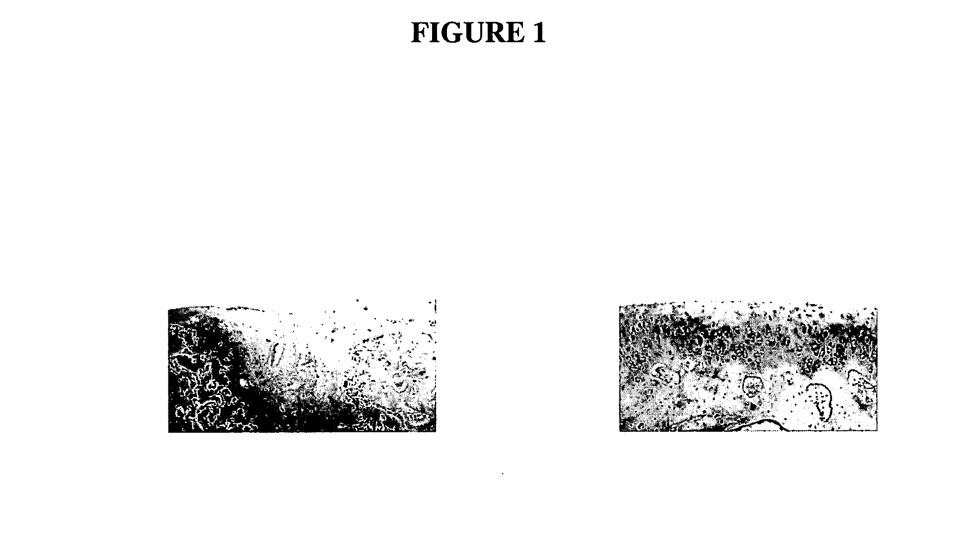
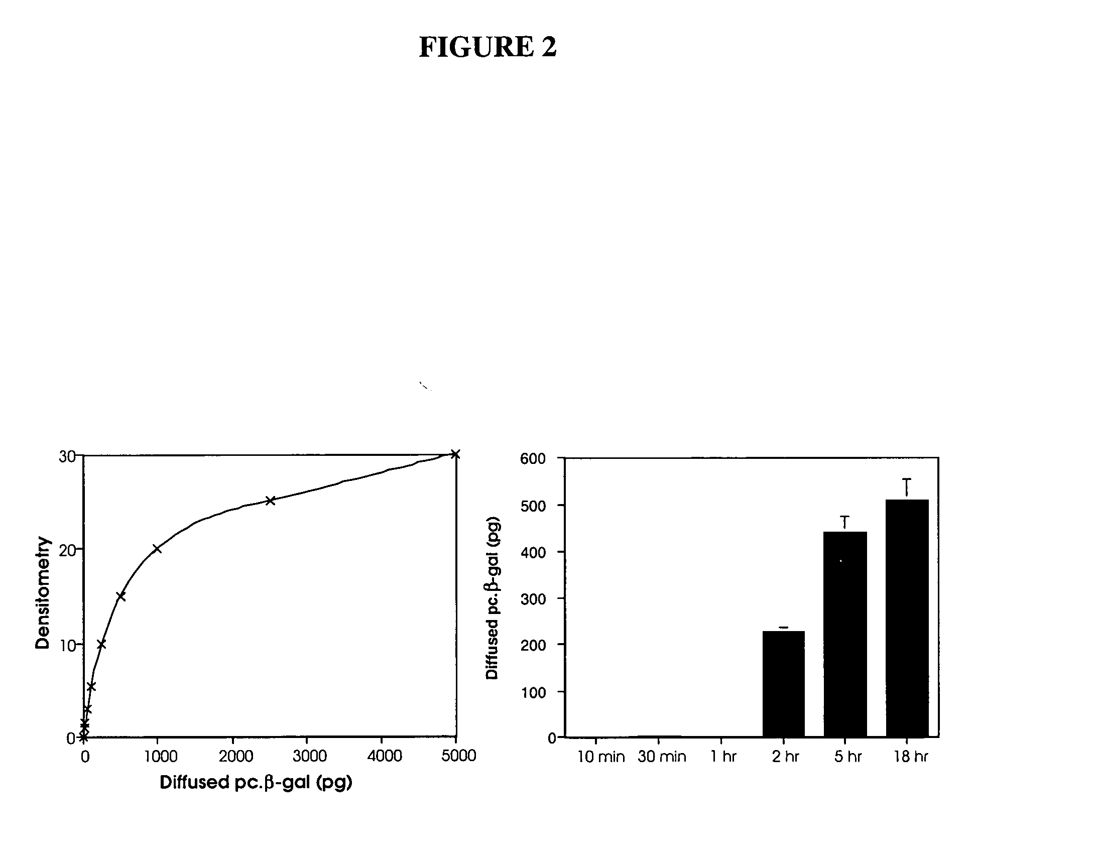
[0147] In our preliminary in vivo study with marker gene plasmid DNA in a rabbit full-thickness defect model, we detected marker protein in many mesenchymal cells under the defect area 1 wk postsurgery. It is clear that these cells take up the naked DNA plasmid and express protein. This study characterizes the duration of active expression of the naked plasmid DNA. 70 μg of pc.βgal / collagen or collagen sponge with control plasmid are implanted in full-thickness articular cartilage defect in 12 adult (9-month-old) male New Zealand White rabbits and samples analyzed after 3, 7, 10, and 21 days postimplantation (3 rabbits=6 knees per time point). These time points are based on other studies using naked plasmid DNA for regional gene therapy that showed active plasmid for up to 3 weeks [54-59]. At each time point, triplicate samples are immunostained with murine monoclonal antibody against β-gal and un...

example 3

Regional Gene Therapy Using Naked Plasmid DNA Encoding the BMP-2 Gene to Promote Hyaline-Like Articular Cartilage Repair

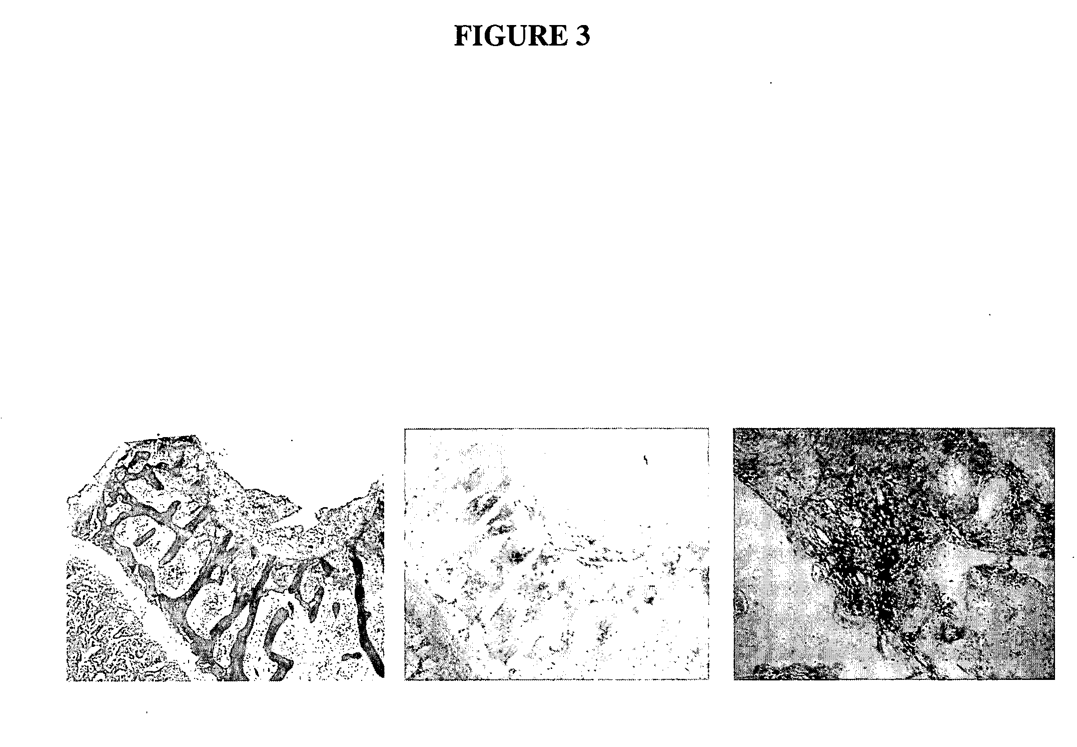
[0151] To document with statistical significance (p<0.05; power=80%) the efficacy of this technique, 9 knees per group are performed. In this study, we examine the cartilage repair at 3 months (18 animals=36 knees) postsurgery in four treatment groups: sponge only, sponge with 6 μg of recombinant human BMP-2, sponge with control plasmid pc.DNA3 (+), and sponge with naked plasmid DNA encoding BMP-2. Repair samples are examined by gross examination, histology (H&E staining), Safranin O staining for proteoglycans, immunostaining (with antibodies to type II collagen and cartilage oligomeric matrix protein).

[0152] Evaluation of cartilage repair in the defects: Cartilage defects are examined macroscopically at sacrifice. Similarity of appearance of the repair cartilage to that of normal adjacent cartilage is assessed based on color, texture, and shape of cartilage surf...


PUM

PropertyMeasurementUnit
Biodegradabilityaaaaaaaaaa

Abstract

The present invention relates to the application of nucleic acid therapy for the repair and regeneration of cartilage. The invention encompasses the introduction of naked DNA encoding bioactive agent(s), whose expression stimulates and otherwise facilitates the repair and regeneration of cartilage. The present invention provides DNA constructs for introduction to the site of cartilage damage. Pharmaceutical compositions comprising nucleic acid encoding one or more bioactive factor, optionally with a matrix or polymer, are provided. Methods for expression of bioactive agent(s) are provided. Methods for enhancing cartilage repair and / or regeneration comprising introduction of bioactive factors as naked DNA are further provided. Methods for the treatment or prevention of cartilage damage in various orthopaedic and rheumatologic conditions are provided.

Description

CROSS-REFERENCE TO RELATED APPLICATIONS [0001] The present application claims the benefit of priority pursuant to 35 U.S.C. § 119 to U.S. Provisional Application 60 / 485,669 filed Jul. 8, 2003, which is incorporated herein by reference in its entirety.FIELD OF THE INVENTION [0002] The present invention relates generally to the application of nucleic acid therapy for the repair and regeneration of cartilage. The invention encompasses the introduction of naked DNA encoding bioactive agent(s), whose expression stimulates and otherwise facilitates the repair and regeneration of cartilage. Methods for the repair and / or regeneration of cartilage for treatment or prevention of cartilage damage in various orthopaedic and rheumatologic conditions are provided. BACKGROUND OF THE INVENTION [0003] More than $215 billion is annually spent on treatment of musculoskeletal conditions in the United States [21]. Arthritis affects almost 1 out of every 8 Americans—about 32 million persons—with an annua...

Claims


Application Information

Patent Timeline
IPC IPC(8): A61KA61K9/14A61K31/70A61K48/00C12N15/74
CPCA61K31/70A61K48/005A61K48/0016
Inventor DICESARE, PAUL
Owner NEW YORK SCHOOL OF MEDICINE
Learn More