Method for identification of proteins from intracellular bacteria

Inactive Publication Date: 2005-10-27
SHAW ALLAN +1
View PDF0 Cites 30 Cited by
  • Summary
  • Abstract
  • Description
  • Claims
  • Application Information

AI Technical Summary

Benefits of technology

[0043] By the term secreted effector protein is meant any protein, which is secreted by the bacteria into the host cell cytoplasm or any intracellular organelle. Secreted effector protein may have great influence on the host/pathogen relation and due to its presence in the host cell cytoplasm may be targeted to the proteasome and presented as MHC-class I antigens on the surface of the host cell. Secreted effector proteins may be secreted by one of several Sec-dependent or independent systems (e.g. Type I, Type II, Type III, Type IV) described in the literature.
[0044] Any bacteria, which has the ability to infect and propagate inside a eukaryotic host cell (e.g. Chlamydia, Salmonella, Shigella, Listeria, Legionella, Yersinia). The definition includes intracellular bacteria, which are obligate intracellular meaning that they may only live and propagate using an eukaryotic host cell or facultative intracellular meaning that they may both sur

Problems solved by technology

If left untreated Chlamydia infections may become chronic with severe complications such as sterility, blindness and potentially thrombosis.
Due to the intracellular developmental cycle persistent Chlamydia infections may cause an aberrant immune response, which fails to clear the organisms.
However, none of these candidates have been proven efficient in vaccine trials.
Type III secreted proteins, however, lack recognisable signal peptidase cleavage sites and no consensus sequence for proteins secreted by this system in Chlamydia has been recognized, such may be restricted to the particular organism in question.
Drawbacks of such a screening is the eukaryotic expression of bacterial proteins which may differ from bacterial expression in a way that alters the probability of processing in the proteasome and the very

Method used


Image

  • Method for identification of proteins from intracellular bacteria
  • Method for identification of proteins from intracellular bacteria
  • Method for identification of proteins from intracellular bacteria

Examples


Example

Example 1

Infection of Mammalian Cell Cultures

[0184] Semi-confluent HeLa, HEp-2 or McCoy (ATCC, Rockville, Md., USA) cell monolayers were infected with one inclusion forming unit (IFU) of C. pneumoniae VR1310, C. trachomatis serovar A (HAR-13), D (UW-3 / Cx) or L2.(434 / Bu)(ATCC) as previously described in [19.] and [17.] The infection medium consisted of RPMI 1640, 25 mM HEPES, 10% FCS. 1% w / v glutamine, 10 mg / ml gentamycin for C. trachomatis A and D and RPMI 1640, 25 mM HEPES, 5% FCS.1% w / v glutamine, 10 mg / ml gentamycin for C. trachomatis L2.

Example

Example 2

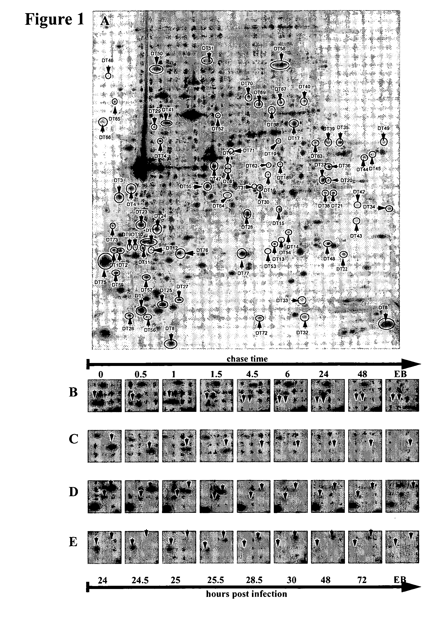
Pulse Labelling / Chase

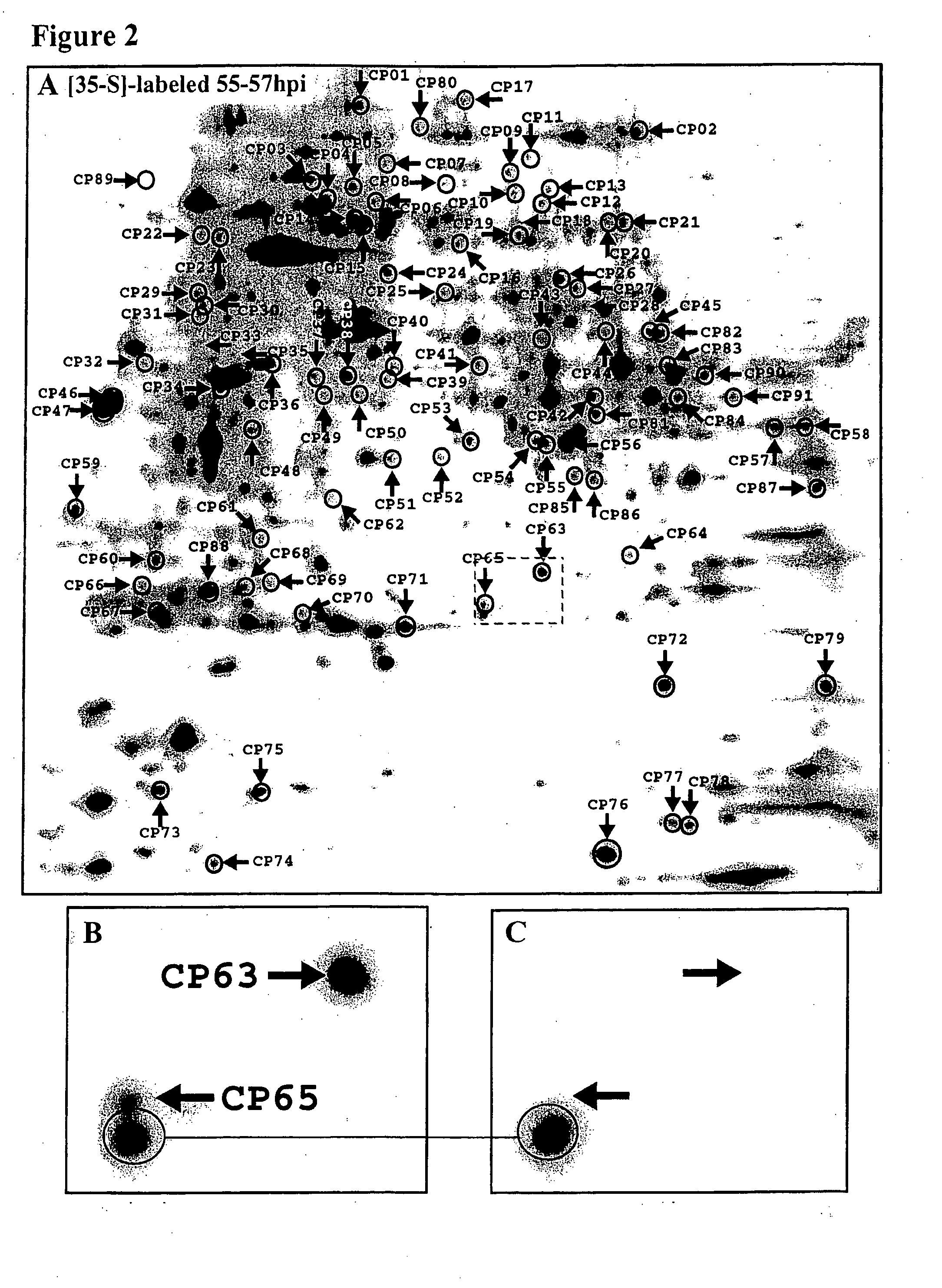
[0185] To label chlamydial proteins for two hour periods, infected cell cultures were incubated in a medium containing RPMI 1640, 10 mg / ml gentamycin, 40 μg / ml cycloheximide, 100 μCi / ml [35S]-methionine / cysteine (Promix, Amersham Pharmacia Biotech, Uppsala, Sweden) as described previously (Shaw et al., 1999,2000) [18.] [19.]. After labelling the labelling medium was changed to normal growth medium following two washes in normal growth medium and the infected cells were harvested at different points in time after labelling. Similarly, labelled EB proteins were obtained by allowing the Chlamydia to grow until 72 h.p.i. after the two hour labelling periods. The labelled EB were then harvested and purified using two consecutive steps of density gradient ultracentrifugation essentially as described for C. trachomatis in (Schacter and Wyrick, 1994) [22.]and for C. pneumoniae (Knudsen et al. 1999 [17.]). Proteins in the EB preparation and pulse chase prepar...

Example

Example 3

Sample Preparation

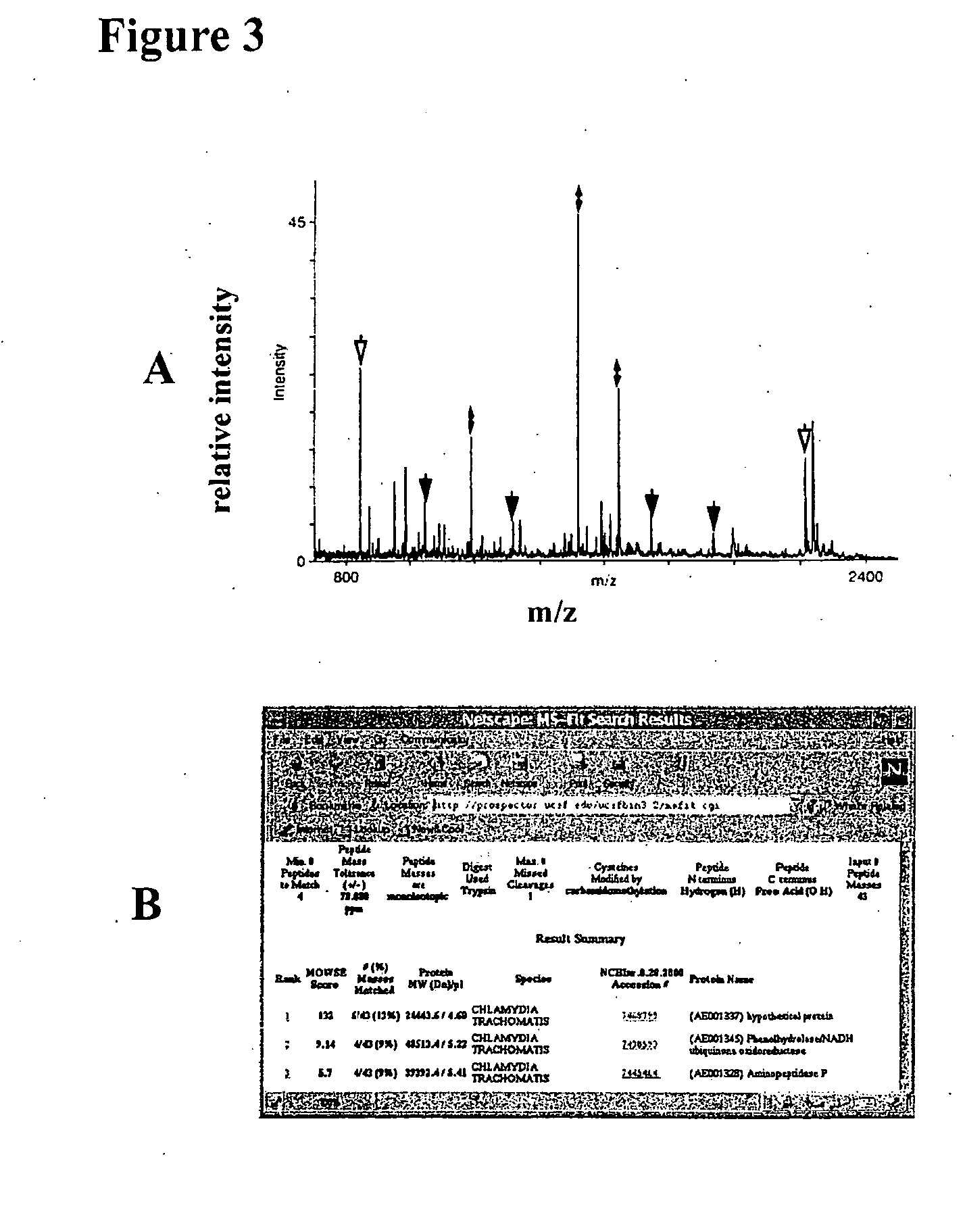
[0186] Following [35S]-labelling cells were washed twice in PBS and solubilised in a standard lysis buffer containing 9 M Urea, 4% w / v 3-[(3-cholamidopropyl)dimethylammonium]-1-propanesulfonate (CHAPS; Roche, Germany), 40 mM Tris Base, 65 mM DTE and Pharmalyte 3-10 (Amersham Pharmacia Biotech). For the enrichment of high molecular weight and hydrophobic proteins 7 M urea, 2 M thiourea, 4% w / v 3-[(3-cholamidopropyl)dimethylammonium]-1-propanesulfonate (CHAPS; Boehringer Mannheim, Germany), 40 mM Tris Base,65 mM dithioerythretiol (DTE) and 2% vol / vol Pharmalyte 3-10 (Amersham Pharmacia Biotech) was used essentially according to (Harder et al. 1999 [23.]). Samples containing whole cell lysates or purified EB were sonicated and centrifuged at 10 000×g for 10 min. Samples were stored at −70° until used.


PUM

PropertyMeasurementUnit
Fractionaaaaaaaaaa
Fractionaaaaaaaaaa
Fractionaaaaaaaaaa

Abstract

The present invention relates to a novel combination of methods that enables identification of proteins secreted from intracellular bacteria regardless of the secretion pathway. The invention further provides proteins that are identified by these methods. Secreted proteins are known to be suitable candidates for inclusion in immunogenic compositions and/or diagnostic purposes. The invention also provides peptide epitopes (T-cell epitopes) from the identified secreted proteins, as well as nucleic acid compounds that encode the proteins. The invention further comprises various applications of the proteins or fragments thereof, such as pharmaceutical and diagnostic applications.

Description

TECHNICAL FIELD OF THE INVENTION [0001] The present invention relates to a novel combination of methods that enables identification of proteins secreted from intracellular bacteria regardless of the secretion pathway. The invention further provides proteins that are identified by these methods. Secreted proteins are known to be suitable candidates for inclusion in immunogenic compositions and / or diagnostic purposes. The invention also provides peptide epitopes (T-cell epitopes) from the identified secreted proteins, as well as nucleic acid compounds that encode the proteins. The invention further comprises various applications of the proteins or fragments thereof, such as pharmaceutical and diagnostic applications. BACKGROUND OF THE INVENTION [0002] The Chlamydia are obligate intracellular bacteria, which multiply inside eukaryotic host cells and are important human pathogens. The order Chlamydiales comprises one family (Chlamydiaceae) containing one genus (Chlamydia), which is divi...

Claims


Application Information

Patent Timeline
IPC IPC(8): A61K39/00C07K14/295G01N33/68
CPCA61K39/00A61K2039/505A61K2039/53G01N2550/00G01N33/6803G01N33/6878G01N2333/81C07K14/295
Inventor SHAW, ALLANVANDAHL, BRIAN
Owner SHAW ALLAN
Why Eureka
Try Eureka