Method of altering the binding specificity of proteins by oxidation-reduction reactions

a technology of oxidation reduction reaction and binding specificity, which is applied in the field of altering the binding specificity of proteins by oxidation reduction reaction, can solve the problems of difficult collection of large amounts of autoantibodies for commercial use, limited amount of such blood that can be obtained from phlebotomy of individual patients or pooling blood from a group of patients known to test positive for autoantibodies, and other methods of obtaining autoantibodies, such as screening phage libraries

Active Publication Date: 2005-11-24
REDOX REACTIVE REAGENTS LLC
View PDF10 Cites 27 Cited by
  • Summary
  • Abstract
  • Description
  • Claims
  • Application Information

AI Technical Summary

Benefits of technology

[0081] Regarding each of the Examples 1-17 described herein, unless otherwise noted, the following procedure was typically used: A 10 ml sample of whole blood or 5 ml of serum or plasma from a normal aPL-negative subject and 4-5 ml of packed mammalian red blood cells were added to a vial containing 30 ml of Biomerieux brand bacterial culture growth medium (containing at least the following ingredients: distilled water, soybean-casein digest broth, yeast extract; dextrose; sucrose; hemin; menadione (vitamin K3); pyridoxal HCl (vitamin B6); and sodium polyanetholesulfonate (SPS) and charcoal. Then, the mixture was incubated, with rocking or shaking, at 37° C. for a period of 18-22 hours. Following the incubation and centrifugation, a sample of the incubated blood or serum / RBC was tested for the presence of antiphospholipid antibodies (aPL) using a comprehensive in-house ELISA aPL format that provides 24 separate aPL test results. The testing procedure is described in greater detail in the following publications, incorporated herein by reference: Wagenknecht D R, et al., The Evolution, Evaluation and Interpretation of Antiphospholipid Antibody Assays, Clinical Immunology Newsletter, Vol. 15, No. 2 / 3 (1995) pp. 28-38 and McIntyre J A, et al., Frequency and Specificities of Antiphospholipid Antibodies (aPL) in Volunteer Blood Donors, Immunobiology 207(1): 59-63, 2003.
[0082]FIG. 1 shows the 24 separate aPL specificities that were tested for by using the comprehensive in-house ELISA aPL format. Four specificities were assessed, 1) aPS=antiphosphatidylserine, 2) aCL=anticardiolipin, 3) aPE=antiphosphatidylethanolamine, and 4) aPC=antiphosphatidylcholine. For each of these aPL specificities, three immunoglobulin isotypes were sought, IgG, IgA and IgM. Each specificity and each isotype were assessed in the presence (dependent) and absence (independent) of a buffer diluent supplement, 10% adult bovine plasma (ABP), which contains the phospholipid-binding plasma proteins) or 1% bovine serum albumin, (BSA, which is devoid of phospholipid-binding plasma proteins), respectively. The final dilution of the subjects' blood samples was between 1 / 50 and 1 / 100.
[0083] The results in the 24 aPL specificities obtained for the various experiments described herein are given in the accompanying figures. The positive / negative findings are expressed in multiples of the means (MoM) based on testing plasma samples from 775 normal blood donors, as described in McIntyre J A, Immunobiology, above. The presence of +++ indicates strong antibody activity. The markers of + and ++ indicate low and intermediate antibody activity, respectively. The figures also provide the normal range values for each aPL specificity and isotype combination.
[0084] A positive result in the column indicated as PL binding protein “dependent” means that the antiphospholipid antibody (aPL) is actually binding to a plasma protein that initially has bound to the particular phospholipid indicated. Plasma proteins that typically can be bound by PS and CL include the following: beta2-glycoprotein I, prothrombin, protein C, protein S, annexin V, and complement components Factor H and C4 (see, for example, McIntyre, J. A., Wagenknecht, D. R. and Faulk, W. P. Antiphospholipid antibodies: Discovery, definition, detection and disease. Prog. Lipid Res. 42(3): 176-237, at page 182). The physiological nature of the plasma protein binding is not known precisely for all of the phospholipids, but such binding is thought to induce conformational changes in the plasma protein structure, thereby exposing novel or cryptic epitopes that then are targeted by the individuals' autoantibodies. Plasma proteins that typically can be bound by phospholipid PE include the following: high and low molecular weight kininogens, and factor XI and prekallikrein. The latter two proteins can be detected by virtue of their fidelity in binding to high molecular weight kininogen. The plasma proteins that bind to PC have not yet been defined. In certain experiments, plasma-protein independent aPL are observed (see FIG. 3). A possible explanation for this activity is that it represents the presence of residual phospholipid-binding plasma proteins that are present in the original blood sample.

Problems solved by technology

Previously, collection of large amounts of autoantibodies for commercial use has been difficult because it was thought that the autoantibodies had to be obtained from individuals having an autoimmune disease or testing positive for autoantibodies in standard assays.
The amount of such blood that can be obtained from phlebotomy of individual patients or by pooling blood from a group of patients known to test positive for autoantibodies is limited.
Other methods of obtaining autoantibodies, such as screening phage libraries as described in U.S. Pat. No. 5,885,793, may be difficult and time-consuming.

Method used


Image

  • Method of altering the binding specificity of proteins by oxidation-reduction reactions
  • Method of altering the binding specificity of proteins by oxidation-reduction reactions
  • Method of altering the binding specificity of proteins by oxidation-reduction reactions

Examples


example 1

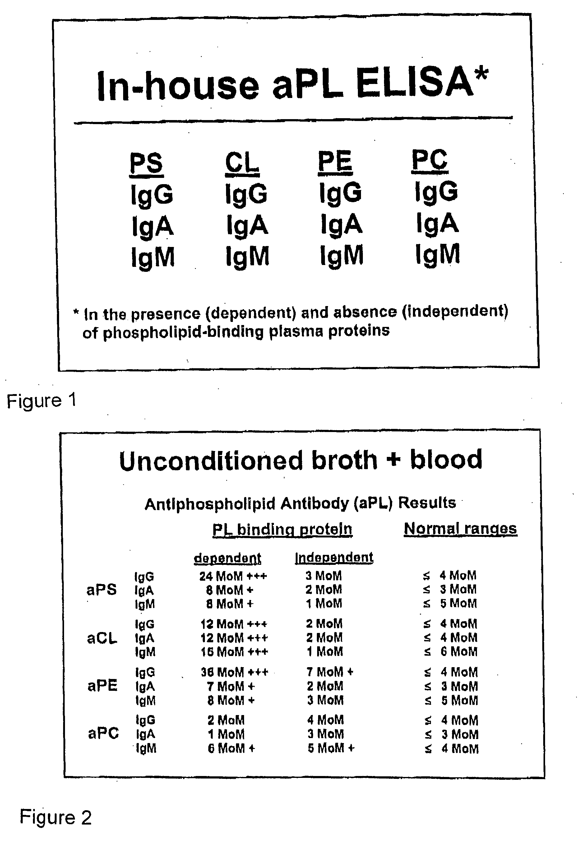
[0085] A sample of blood from a normal subject was incubated and tested according to the procedure described above. The results of the aPL ELISA are shown in FIG. 2. As shown in FIG. 2, the incubated blood sample shows a dramatic presence of autoantibody activity, in comparison to the normal, untreated blood shown in the Normal ranges column. In particular, strong autoantibody activity is shown in the protein-dependent category for aPS (IgG), aCL (all isotypes), and aPE (IgG). The low or absent IgG aPC autoantibody activity was a characteristic finding in the early examples and in procedures in which hemin was used as the oxidizing agent. This result indicates that autoantibodies to PC, especially of the IgG isotype are different and perhaps do not become activated in the same way as do the others. In later experiments, it was found that significant levels of aPC can be detected in samples that were treated with KMnO4 (data not shown).

example 2

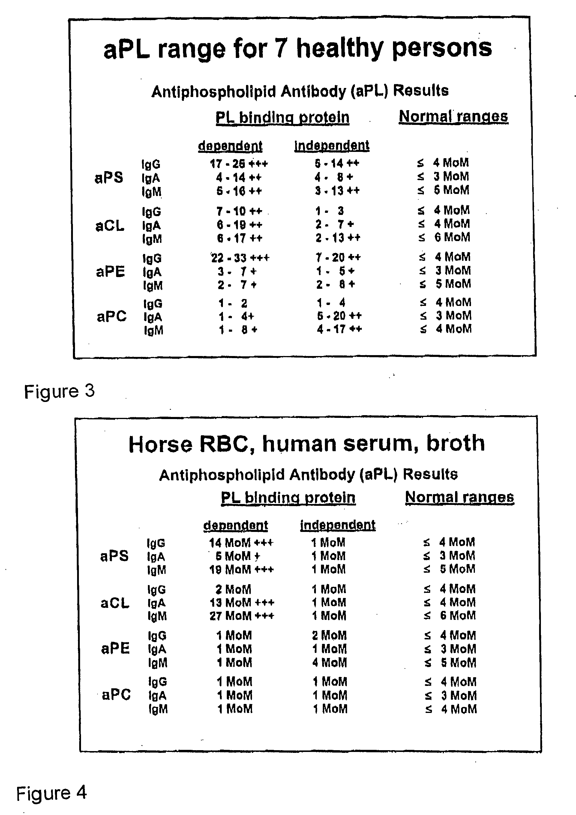
[0086] Blood samples drawn from seven healthy subjects were incubated and tested according to the procedure described above. In particular, all seven subjects' bloods were drawn within a 20 minute period and incubated for 20 hours in identical conditions. FIG. 3 is a composite table showing the range of aPL seroconversion for these seven samples. These results show that there are variations in the aPL levels detected as well as the isotypes present among different individuals. Nevertheless, as shown by the invention, each individual had aPL antibodies that could be detected after incubation.

example 3

[0087] In a first experiment, a serum sample from a normal subject was incubated and tested according to the basic procedure described above. In the incubation mixture, horse red blood cells (RBC) were used instead of human RBC. The results of the aPL ELISA are shown in FIG. 4. As shown in FIG. 4, significant aPL activity was obtained, particularly with respect to aPS (IgG and IgM) and aCL (IgA and IgM).

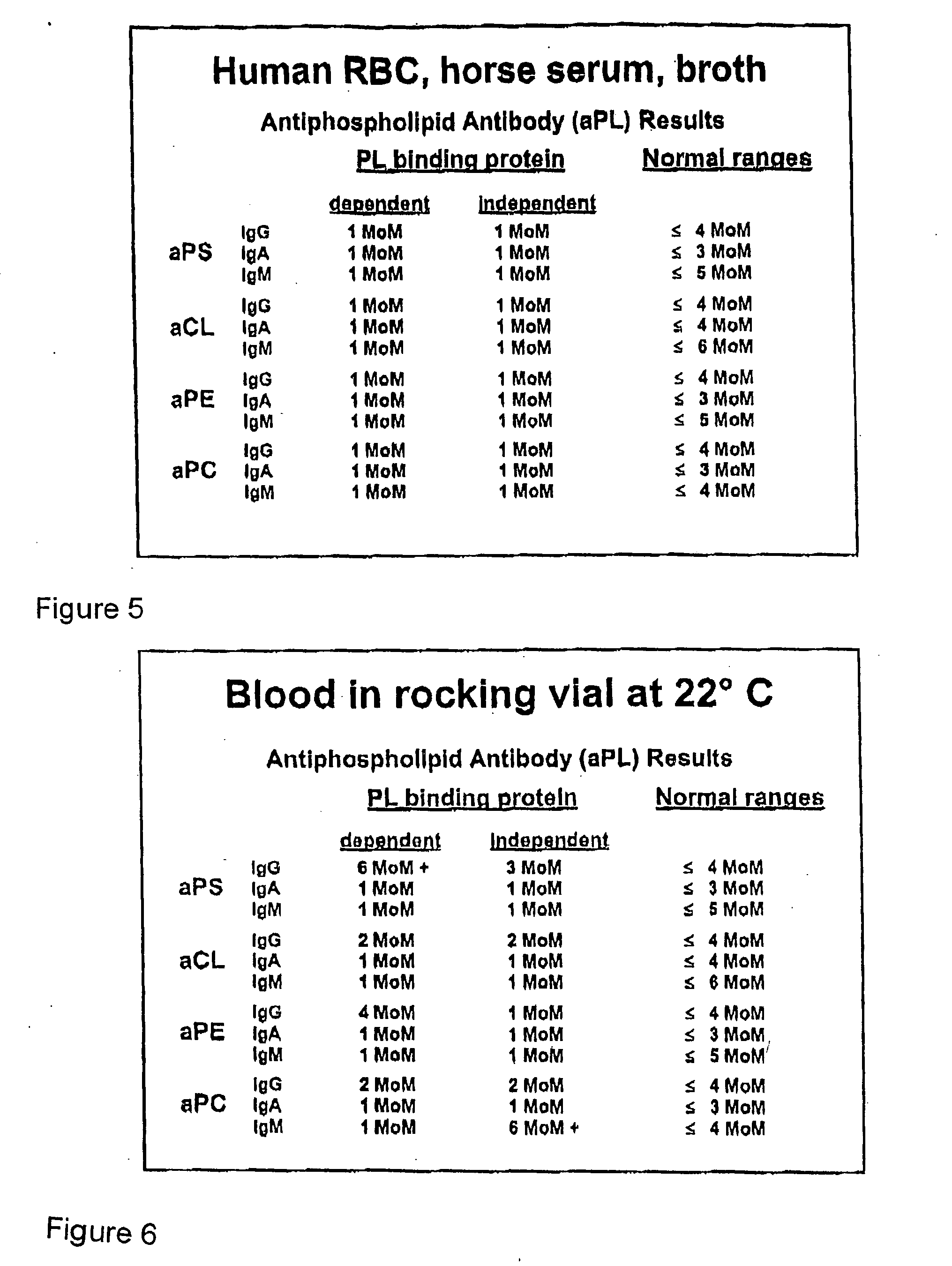
[0088] In a second experiment, a horse serum, instead of human serum, was incubated with human RBC and tested according to the basic procedure described above. The results of the aPL ELISA are shown in FIG. 5. As shown in FIG. 5, aPL activity was not obtained. (The ELISA assay used in this experiment utilized human-antibody-specific alkaline phosphatase labeled antibody probes to detect aPL, so whether the incubated sample contained horse aPL is unknown.)

[0089] The results shown summarized in FIGS. 4 and 5 unequivocally demonstrate that all aPL that are obtained during the seroconv...


PUM

PropertyMeasurementUnit
temperatureaaaaaaaaaa
electric potentialaaaaaaaaaa
conformationalaaaaaaaaaa

Abstract

The binding specificity of at least one protein suspended or dissolved in a liquid medium is reversibly altered by exposing the protein to an oxidizing agent or an electric current. A masked protein such as an autoantibody can be detected, isolated and recovered from a biological fluid by subjecting the biological fluid to an oxidizing agent or an electric current to change the binding specificity of masked proteins contained therein.

Description

[0001] The present application is a continuation-in-part of U.S. patent application Ser. No. 10 / 863,365, filed on Jun. 9, 2004, which application claimed the benefit of the filing date of U.S. Provisional Application No. 60 / 476,607, filed Jun. 9, 2003. These applications are incorporated herein by reference.FIELD OF INVENTION [0002] The present invention relates to a method of altering a binding specificity of a protein that has a binding specificity that can be altered by oxidation-reduction reactions. The present invention further relates to a method of reversibly altering the binding specificity of antibodies, particularly masked autoantibodies that are naturally present in the blood, plasma, serum or other bodily fluids of normal subjects. BACKGROUND OF THE INVENTION [0003] The term “autoimmune disease” refers to a group of diseases wherein the immune system mistakenly attacks cells, tissues and organs of a person's own body. Typically, autoimmune diseases involve antibody bindi...

Claims


Application Information

Patent Timeline
Patent Type & Authority Applications(United States)
IPC IPC(8): C07K16/18C12N15/01G01N33/53
CPCC07K16/00C07K16/18C07K2317/21G01N33/6854G01N33/5375G01N33/564G01N33/5306
Inventor MCINTYRE, JOHN A.
Owner REDOX REACTIVE REAGENTS LLC
Why Eureka
Try Eureka