Liposome-containing radiographic contrast medium and preparation method thereof

a radiographic contrast medium and liposome technology, applied in the direction of dispersed delivery, pharmaceutical delivery mechanism, therapy, etc., can solve the problems of difficult to obtain high contrast images, inapplicability of foregoing methods, and inability to detail examination of tissue or disease regions, etc., to achieve efficient and stable confinement, enhance delivery efficiency and selectivity

Inactive Publication Date: 2006-01-26
KONICA MINOLTA MEDICAL & GRAPHICS INC
View PDF6 Cites 4 Cited by
  • Summary
  • Abstract
  • Description
  • Claims
  • Application Information

AI Technical Summary

Benefits of technology

[0009] It is therefore an object of the present invention to provide a radiographic contrast medium exhibiting enhanced delivery efficiency and selectivity by enclosing contrast medium material in liposome vesicles.
[0010] It is a further object of the invention to provide a radiographic contrast medium containing a liposome and a preparation method thereof in which an iodine compound is enclosed efficiently and stably without use of toxic organic solvents and a preparation method thereof.

Problems solved by technology

On the contrary, the difference in X-ray absorption between different soft tissues is relatively small, making it difficult to obtain high contrast images.
However, the foregoing compounds are promptly discharged from the lumen region without interacting with tissue or disease regions, which is not useful for detailed examination of the tissue or disease region, specifically such as cancer tissue.
Accordingly, the foregoing methods were not practically applicable due to toxicity of retained solvents.
Although water-soluble electrolytic chemicals can be included in a liposome through interaction of a charge of the chemicals with that of a charged lipid, such a means is not applicable to water-soluble non-electrolytic chemicals.
It has been generally desired to allow non-ionic iodine compounds substantially exhibiting low toxicity to be included in a liposome rather than ionic contrast medium compounds, which is not easy from the foregoing reasons.
In these means, however, organic solvents are used, producing problems of safety.
However, although examples of a water-soluble electrolyte are shown therein as a medicinal component, it is unclear whether a water-soluble non-electrolyte is efficiently included in a liposome using this method.
Further, this method is desired to use auxiliary solvents such as ethanol to enhance the enclosure ratio, rendering it difficult to prepare a liposome of a relatively high enclosure ratio without the use of organic solvents.
Even if inclusion of a contrast medium material is done well, problems such as its leaking-out over an elapse of time or the situation of the liposome itself becoming unstable must be taken into account.

Method used


Image

  • Liposome-containing radiographic contrast medium and preparation method thereof

Examples


example 1

Determination of Iodine of Iodine Compound

[0091] A sample (liposome dispersion) was dialyzed with an isotonic saline solution and after completion of dialysis, ethanol was added thereto to destroy the liposome and the quantity of the iodine compound included in the liposome was determined by absorptiometry. The ratio of the foregoing quantity to the total amount of the iodine compound of the sample was represented as an enclosure ratio (wt %).

Preparation of Contrast Medium

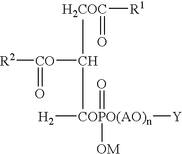
[0092] A mixture of 86 mg of dipalmitoylphosphatidylcholine (DPPC), 38.4 mg of cholesterol and 19.2 mg of PEG-phospholipid (SUNBRIGHT DSPE-020CN, lipid modified with polyethylene glycol, product by NIPPON OIL &FATS CO., LTD.) were added into a stainless steel autoclave and heated with maintaining the autoclave at 60° C., then, 13 g of liquid carbon dioxide was added thereto. While stirring, the pressure within the autoclave was increased from 50 kg / cm2 to 120 kg / cm2 by decreasing the internal volume of the aut...

example 2

[0095] Samples 2-1 to 2-8 were prepared similarly to Sample 1-1 of Example 1, provided that the contrast medium solution was varied with respect to amount or kind, as shown in Table 2.

[0096] The thus prepared samples were measured with respect to the iodine content, using a spectrophotometer to determine the enclosure ratio.

TABLE 2IodineEnclosureSampleContrast MediumContentRatioNo.Solution(mg / ml)(wt %)2-1Iopamidol solution150192-2Iopamidol solution250182-3Iopamidol solution300152-4Iopromide solution240162-5Iohexol solution180182-6Iohexol solution300142-7Iomeprol solution300132-8Ioxilan solution30014

example 3

Determination of Liposome Vesicle Size

[0097] Liposome vesicle size was determined in the following manner. A dispersion containing liposome vesicles enclosing an iodine compound was frozen and fractured, following which carbon was vapor-deposited onto the fractured interfaces and the deposited carbon was observed by an electron microscope (freeze fracture TEM method). The particle size was defined as an arithmetic average of the sizes of 20 liposome particles observed.

Preparation of Contrast Medium

[0098] A mixture of 192 mg of dipalmitoylphosphatidylcholine (DPPC), 76.8 mg of cholesterol and 38.4 mg of PEG-phospholipid (SUNBRIGHT DSPE-020CN, phospholipid modified with polyethylene glycol, product by NIPPON OIL & FATS CO., LTD.) were added into a stainless steel autoclave and heated with maintaining the autoclave at 60° C., then, 13 g of liquid carbon dioxide was added thereto. While stirring, the pressure within the autoclave was increased to 120 kg / cm2 from 50 kg / cm2 by decrea...


PUM


Abstract

A method of pereparing a rediographic contrast medium containing liposomes is disclosed, comprising (a) dissolving a phospholipid and a sterol in a supercritical or subcritical carbon dioxide in the presence of a compound containing a hydroxyl group or a polyalkyleneoxide group, (b) adding thereto an aqueous solution containing an iodine compound to form micelles and (c) discharging the carbon dioxide to form liposomal vesicles enclosing the iodine compound.

Description

[0001] This application claims priority from Japanese Patent Application Nos. JP2004-213290 filed on Jul. 21, 2004; JP2004-238505 filed on Aug. 18, 2004; JP2004-252929 filed on Aug. 31, 2004; and JP2004-252930 filed on Aug. 31, 2004 which are incorporated hereinto by reference. FIELD OF THE INVENTION [0002] The present invention relates to a radiographic contrast medium containing liposome and a preparation method thereof, and in particular to a radiographic contrast medium containing liposome vesicles which are formed by mixing lipid membrane constituents with a supercritical or subcritical carbon dioxide in the presence of at least a compound containing a hydroxyl group or polyalkyleneoxy group and in which nonionic iodine compounds are efficiently and stably enclosed. BACKGROUND OF THE INVENTION [0003] Investigation or diagnosis using X-rays is the nucleus of current medical image diagnosis. So-called hard tissues such as bones and teeth efficiently absorb X-rays and thereby high...

Claims


Application Information

Patent Timeline
Patent Type & Authority Applications(United States)
IPC IPC(8): A61K51/00A61K49/04
CPCA61K49/0466
Inventor UEDA, EIICHINAKAJIMA, AKIHISANAGAIKE, CHIAKIMOTOKUI, YASUYUKI
Owner KONICA MINOLTA MEDICAL & GRAPHICS INC
Why Eureka
Try Eureka