
Methods and agents for treating axonal damage, inhibition of neurotransmitter release and pain transmission, and blocking calcium influx in neurons

a technology of axonal damage and inhibitory neurotransmitter, which is applied in the direction of animal cells, peptide/protein ingredients, depsipeptides, etc., can solve the problems of major health problems, traumatic brain injury is a major health problem, and therapies are unable to repair axonal damage, so as to promote the outgrowth of a mammalian neuron

Inactive Publication Date: 2006-06-29
MT SINAI SCHOOL OF MEDICINE
View PDF6 Cites 0 Cited by
  • Summary
  • Abstract
  • Description
  • Claims
  • Application Information

AI Technical Summary

Benefits of technology

[0015] The present invention is directed towards a method for promoting outgrowth of a mammalian neuron comprising inhibiting the binding of an ankyrin protein of said neuron to an L1-CAM protein, by contacting said neuron with a peptide comprising the amino acid sequence set forth in SEQ ID NO: 2. In one aspect, the present invention is directed towards a

Problems solved by technology

Spinal cord injury affects millions of individuals worldwide, resulting in severe impairment of the physical function of affected persons (e.g., as seen in paraplegia and quadriplegia).
Traumatic brain injury is a major health problem in all developed countries.
Currently available therapies are unable to repair the axonal damage.
), but such studies have not previously been extended to proteins in the presynaptic active zones.
Chronic pain affects millions of individuals; it results in severe impairment of a patient's physical function and is often associated with psychological depression.
These analgesics become less effective over time as patients become sensitized.
Stroke and traumatic head injury result in similar pathological processes in the brain.

Method used


Image

  • Methods and agents for treating axonal damage, inhibition of neurotransmitter release and pain transmission, and blocking calcium influx in neurons
  • Methods and agents for treating axonal damage, inhibition of neurotransmitter release and pain transmission, and blocking calcium influx in neurons
  • Methods and agents for treating axonal damage, inhibition of neurotransmitter release and pain transmission, and blocking calcium influx in neurons

Examples


example 1

Assembly of Expression Constructs

[0093] pL1-CAM: This expression vector contains a nucleotide sequence (FIG. 7; SEQ ID NO: 4) encoding the L1-CAM protein (FIG. 8; SEQ ID NO:5) inserted into the NheI and XhoI sites of pIRES2-EGFP (Clontech). This expression construct provides for the expression of both L1-CAM and enhanced green fluorescent protein (EGFP) from a single bicistronic transcript containing an internal ribosomal entry site (IRES), where expression is regulated by a cytomegalovirus (CMV) promoter.

[0094] The L1-CAM-encoding nucleotide sequence (SEQ ID NO: 4) represents a variant of the nucleotide sequence for rat L1-CAM. The L1-CAM protein (SEQ ID NO: 5) encoded by the nucleotide sequence (SEQ ID NO: 4) is 99.44% identical to the rat L1-CAM protein previously reported by Miura et al. FEBS Lett. 1999;289:91-95 (Genbank Accession # Q05695). The two amino acid sequences differ at only 7 positions (see FIG. 8). Three of these changes are in the 21 amino acid signal sequence, a...

example 2

Expression of Wild-Type and Mutant L1-CAM in In Vitro Cultured Rat Neurons

Materials and Methods

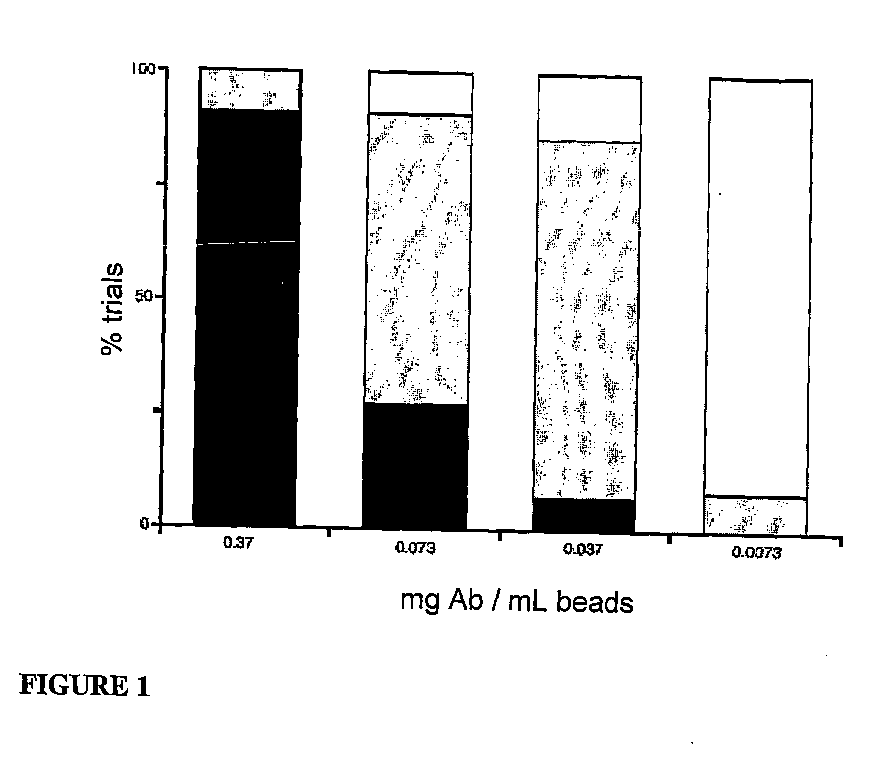
[0105] Cell culture aid transfection: ND-7 cells were generated by poly-ethylene glycol-mediated fusion of N18Tg2 neuroblastoma cells (German Collection of Microorganisms and Cell Cultures, Braunschweig, Germany) with dorsal root ganglion (DRG) cells harvested from neonatal rat. These parent cell lines were cultured in vitro by standard techniques well established in the art (see, e.g., Animal Cell Culture (Freshney, ed.: 1986). ND-7 cells were routinely cultured in L15 medium (Gibco), supplemented with penicillin, streptomycin, L-glutamine, and vitamins (all available from Cell and Molecular Technologies), containing 10% bovine calf serum (Hyclone), and buffered for CO2. Cultured ND-7 cells were plated on sialanized and laminin-coated coverslips 24 hours prior to transfection. Plated cells were transfected with pMyc-L1-CAM, pMyc-L1-CAM-YF or pMyc-L1-CAM-YH expression constructs using Li...

example 3

L1-CAM Cytoskeleton Interactions Depend on L1-CAM Crosslinking,

Material and Methods

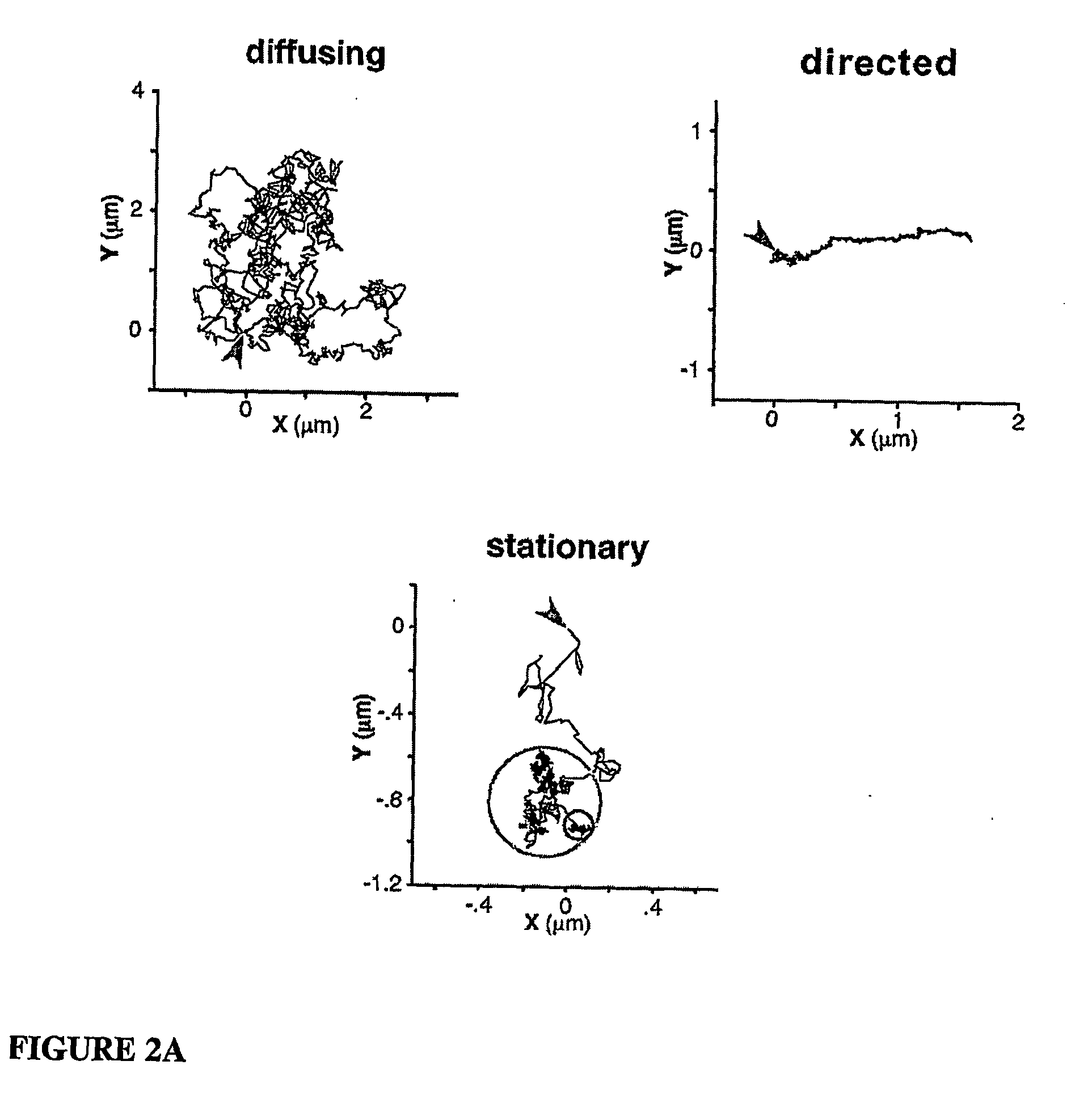
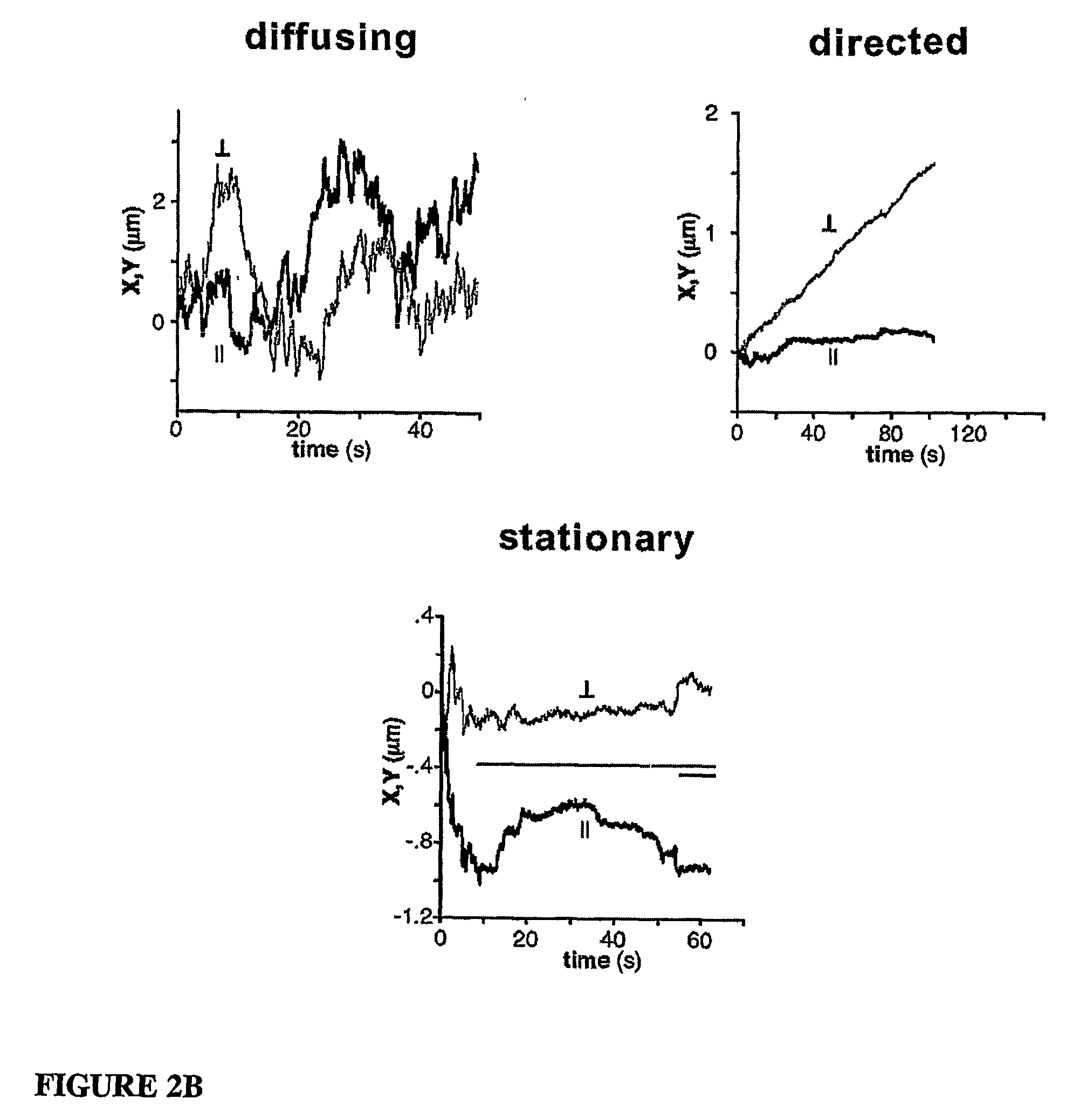
[0110] Cell culture and transfection: ND-7 cells (rat neuroblastoma / DRG hybrid cells) were cultured as described in Example 2. 24 hours prior to transfection, cultured cells were plated on coverslips coated with poly-D-lysine and laminin sealed to the bottom of 35 mm culture dishes (Mattek Corp.). Plated cells were transfected with the pL1-CAM or pMyc-L1-CAM expression constructs using Lipofectamine plus (Invitrogen) according to manufacturer's instructions. Transfected cells were cultured for 24-36 hours, and then used for live microscopy. For laser trap and video microscopy, the medium was replaced with phenol red-free serum-free L15 medium (air buffered) with 20 mM HEPES, 0.1% BSA, 0.5% ovalbumin. Cells transfected with the bicistronic expression constructs express a single mRNA encoding both the myc-L1-CAM protedms and EGFP, such that transfected cells were identified by EGFP fluorescence.

[0111...


PUM

PropertyMeasurementUnit
Cell deathaaaaaaaaaa
Adhesion strengthaaaaaaaaaa
Currentaaaaaaaaaa

Abstract

The present invention pertains to methods to promote outgrowth of, or extension across a substrate of, neuronal cells by inhibiting the interaction between the cytoplasmic tail of the L1-CAM cell surface adhesion molecule and the cytoskeletal protein ankyrin. The invention also pertains to a method to treat diseases characterized by axonal damage such as spinal cord injury, traumatic brain injury, stroke, and neurodegenerative disease by administration of novel peptides that inhibit the binding of the L1-CAM cytoplasmic tail to ankyrin, and to pharmaceutical compositions comprising such peptides. The present invention pertains to the regulation of neuronal signal propagation. Addition of the peptide of the invention disrupts the interaction between the cytoplasmic tail of the cell surface adhesion molecule L1-CAM, the cytoskeletal protein ankyrin, and voltage-gated calcium channels at the presynaptic surface of a neuron leading to a transient redistribution of the calcium channels to cytoplasmic vesicles. This redistribution blocks signal propagation as demonstrated by the inhibition of voltage gated calcium current and a decreased secretion of Substance P, which mediates pain signaling. Furthermore, the invention pertains to a method of blocking calcium flux to protect against neural cell death following stroke or traumatic head injury.

Description

[0001] This application claims priority under 35 U.S.C. §119(e) to U.S. Provisional Applications Ser. Nos. 60 / 480,092 filed Jun. 19, 2003, and 60 / 544,798 filed Feb. 13, 2004 which are incorporated by reference herein in their entirety.[0002] The research leading to this invention was supported, in part, by Grant No. GM63192-01 and Grant No. N537443 awarded by the National Institutes of Health. Accordingly, the United States government may have certain rights to this invention.FIELD OF THE INVENTION [0003] The present invention pertains to methods to promote outgrowth of, or extension across a substrate of neuronal cells. The invention also pertains to methods to block calcium signaling through neurons. BACKGROUND OF THE INVENTION [0004] The adhesion and migration of adherent cells depends on the function of cell-surface glycoproteins that mediate the adhesive contact between the cell surface and molecules in the cellular microenvironment. In quiescent, non-migratory cells, adhesion ...

Claims


Application Information

Patent Timeline
IPC IPC(8): A61K38/17C07K14/47C07H21/04C12P21/06A61K
CPCA61K38/17C07K14/47C07K2319/03
Inventor FELSENFELD, DANDIVERSE-PIERLUISSI, MARIA
Owner MT SINAI SCHOOL OF MEDICINE
Learn More