Visible-near infrared spectroscopy in burn injury assessment

a burn injury and infrared spectroscopy technology, applied in the field of medical devices, can solve the problems of scarring, tissue damage, even death, accurate assessment of burn severity remains a problem for burn specialists, and the ability to distinguish between burns that will heal on their own versus those that will require surgical intervention is particularly challenging

Active Publication Date: 2006-07-13
NAT RES COUNCIL OF CANADA
View PDF17 Cites 193 Cited by
  • Summary
  • Abstract
  • Description
  • Claims
  • Application Information

AI Technical Summary

Problems solved by technology

Cutaneous burns are one of the most destructive insults to the skin, causing damage, scarring and even death of the tissue.
Despite the large number of annual burn cases, the accurate assessment of burn severity remains a problem for the burn specialist.
The ability to distinguish between burns that will heal on their own versus those that will require surgical intervention is particularly challenging.
Highly destructive burns have only a marginal residual blood supply to the dermis that may result in ischemia and ultimately necrosis of the dermis, as the re-epithelialization of the tissue depends on the viable dermis below the burned tissue.
Deep partial and full thickness injuries involve destruction of the dermal layer and what little if any remains of the dermis is insufficient to regenerate due to a reduced dermal blood supply.
However, the major drawback of this technique is that it provides a static picture of the injury reflecting the extend of tissue damage at the time the biopsy was taken.
Since burn injuries are dynamic and change over the early postburn period, a single biopsy taken at the initial assessment of the injury may not accurately predict outcome.
While this method has demonstrated success in distinguishing superficial from full thickness burns (i.e. presence or absence of fluorescence), it cannot easily differentiate those burn types that are between the two extremes.
Furthermore, the extended washout times of the dye limit the frequency with which it can be used to assess a dynamic injury.
As a result, indocyanine green has not yet met with clinical acceptance even though it has been available for burn diagnosis for over a decade.
As can be seen, this process is invasive as it requires the injection of fluorescent dyes and also relies on the use of a database for characterizing the burn injury.

Method used


Image

  • Visible-near infrared spectroscopy in burn injury assessment
  • Visible-near infrared spectroscopy in burn injury assessment
  • Visible-near infrared spectroscopy in burn injury assessment

Examples


example i

Animal Model

[0035] Following a 10 day acclimatization period, adult Yorkshire cross swine weighing between 40 and 50 kg were premedicated with an intramuscular injection of midazolam (0.3 mg / kg), atropine (0.02 mg / kg), and ketamine (20 mg / kg). Anesthesia was then induced by mask and the pigs were intubated and mechanically ventilated. Isoflurane (1.5-2.5%), was delivered through the ventilator (via 40-60% oxygen mixed with medical air at 3.0 Umin) to maintain anesthesia for the duration of the experiment. Systemic oxygen saturation, heart rate, and blood pressure were monitored throughout the experiment. Core body temperature was maintained at 39.0° C.±0.5° C. Blood samples for blood gas and electrolyte analyses were acquired prior to thermal injury and every hour thereafter.

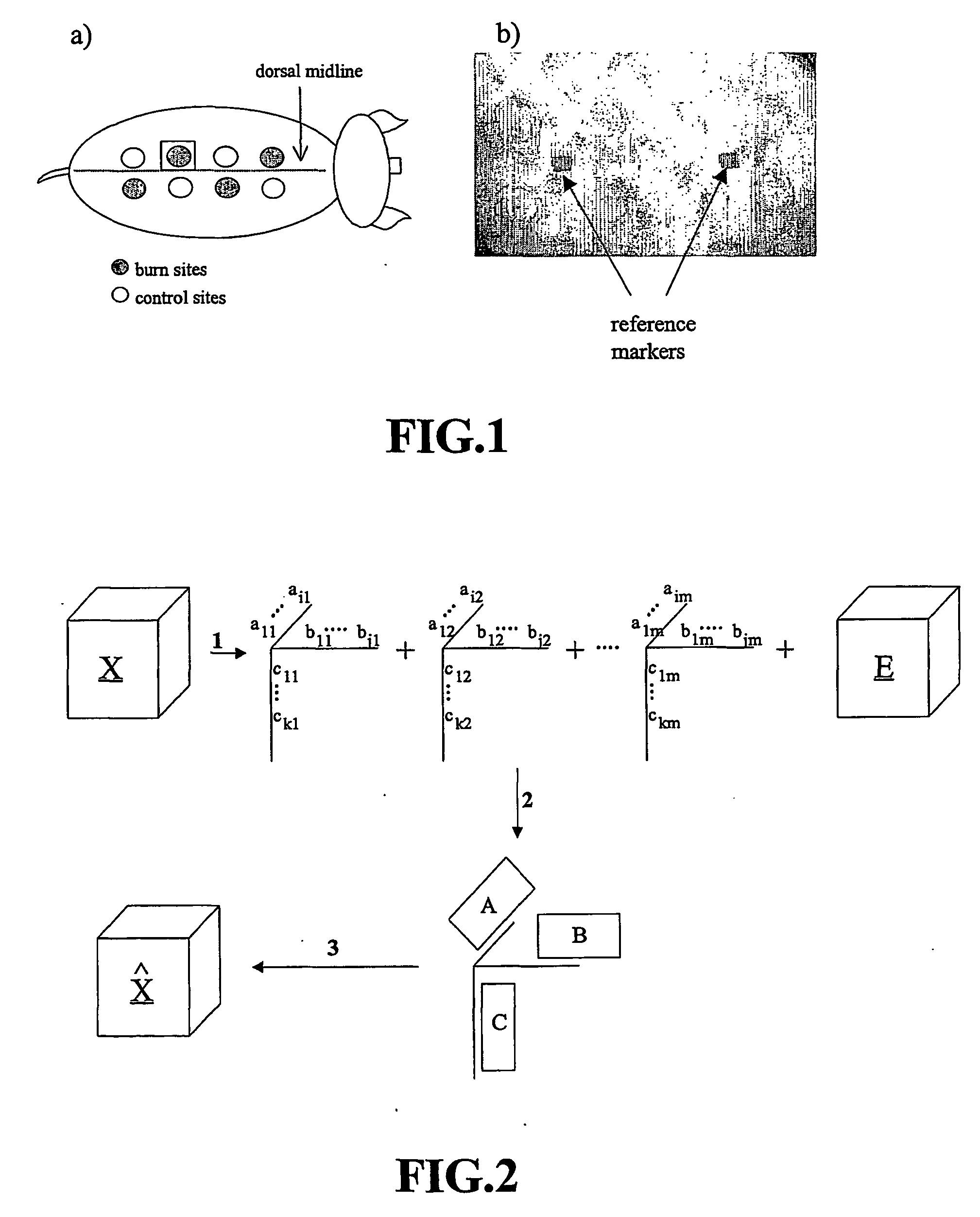
[0036] Following anesthesia, both sides of the dorsal midline were shaved and eight sites, each 3 cm in diameter, were marked on the back of the animal using a custom-made template. Four of the sites were loca...

example ii

Visible-Near Infrared Spectroscopy

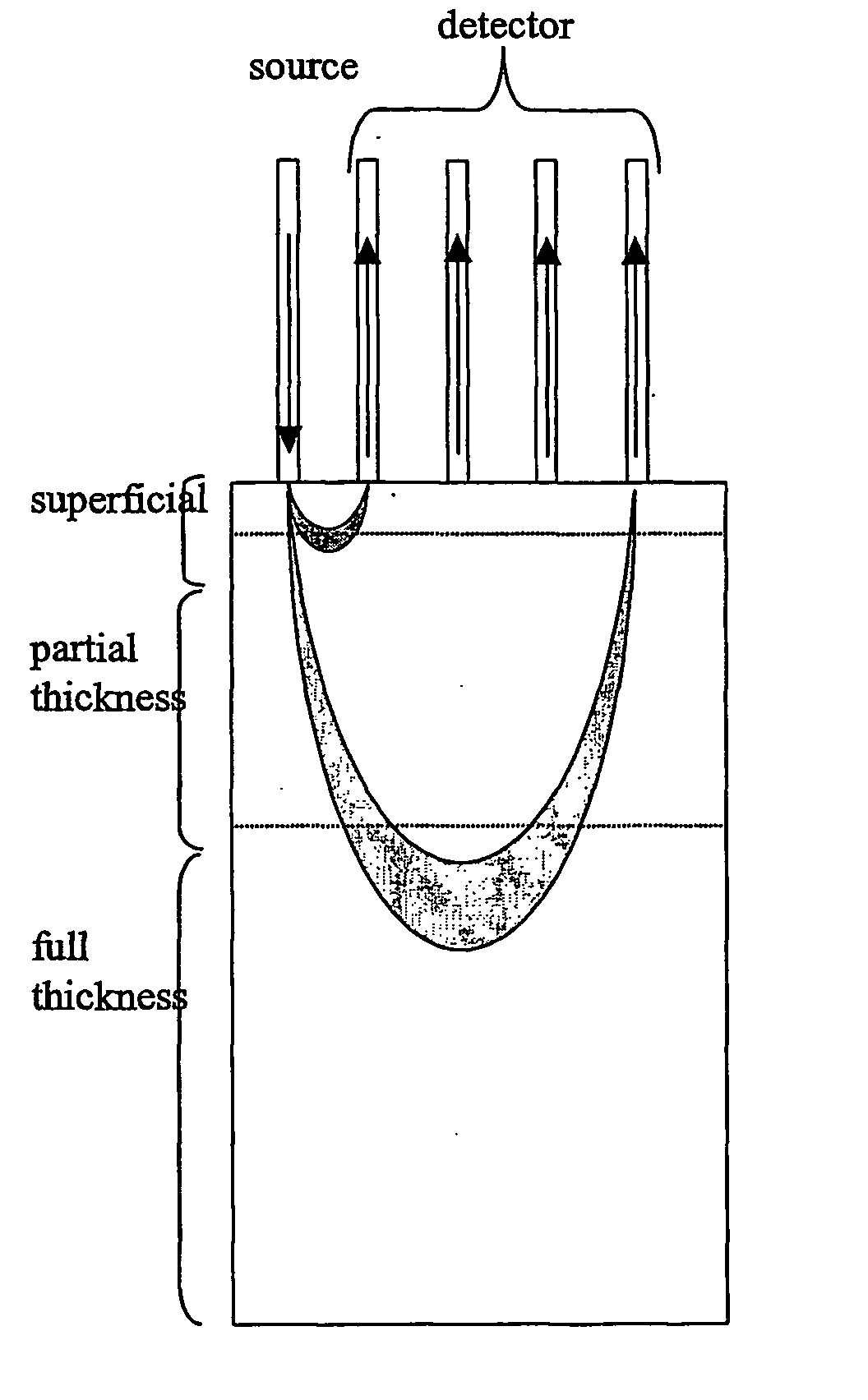
[0039] Visible-near infrared spectra were collected with an NIRSystems 6500 (Foss, Silver Springs, Mass.) spectrometer using a custom bifurcated fiber optic bundle (Fiberguide Industries, Stirling, N.J.). The multifiber probe consisted of five optical fibers, one to illuminate the tissue and four to collected the remitted light. The illumination and collection fibers were 2 m in length with a core diameter of 600 and 200 μm, respectively. The fiber order at the head of the probe were placed in a co-linear arrangement beginning with the illumination and subsequent collection fibers spaced 1.5 mm from each other. Therefore, the distance of the four collection fiber from the illumination source were 1.5, 3, 4.5, and 6 mm. The illumination optical fiber was coupled to a 100 W quartz tungsten halogen white light source (Oriel, Strattford, Conn.). The four collection optical fibers were placed at the entrance of an imaging spectragraph (Sciencetech Inc.,...

example iii

Multivariate Data Processing

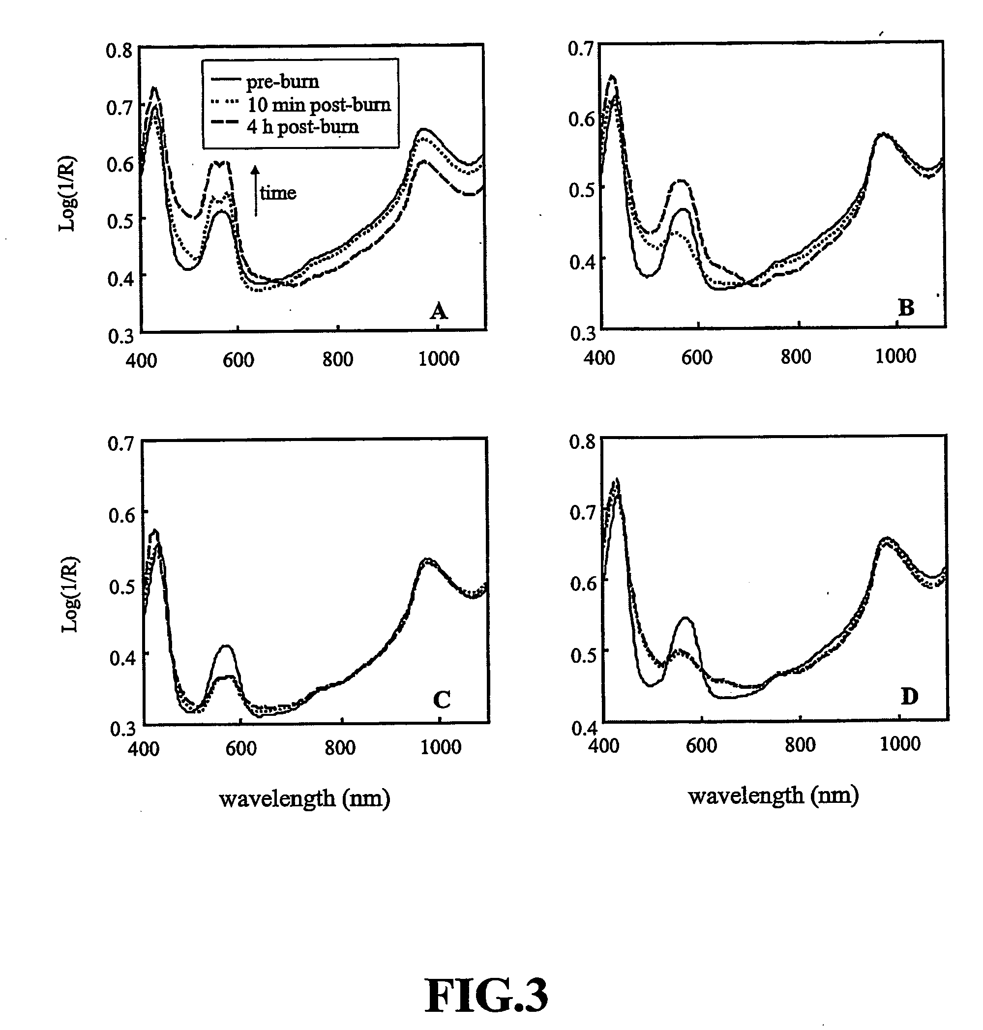
[0045] A time series of near infrared spectra were collected from selected sites on the dorsa of 5 animals. The first time point in the series was acquired prior to thermal injury. Subsequent points in the time series were acquired at fixed time intervals after the injury. The experimental design results in a rich set of data consisting of many variables (reflectance response over the 400-2500 nm wavelength range) observed on several occasions. (longitudinal measurements) and measured at several sites or groups (cross-sectional data) over a sample population of 5 animals. Parallel factor analysis is used to try to isolate and recover the spatial, spectral and temporal changes in reflectance of the skin due to the varying degree of thermal insult.

[0046] Parallel factor analysis is used to explore the time series of spectral data by identifying the “pure” molecular species that contribute to the spectra and determining how these “pure” species are affecte...


PUM


Abstract

A non-invasive method of characterizing burn injuries using near infrared spectroscopy is described. In the method, a beam of light is emitted into the burnt tissue portion at two or more different tissue depths. The spectra are then compared using multivariate analysis to determine diagnostic regions of the spectra. This information is used to categorize the burn. In some cases, the diagnostic regions correspond to wavelengths related to the hemodynamics of the tissue portion. The spectra can also be repeated over time, thereby allowing trends and changes in the spectra to be measured. This data is in turn used to categorize the burn as either a superficial burn, partial thickness burn, deep partial burn or a full thickness burn. Once the burn has been categorized, the clinician can intervene as needed to treat the burn.

Description

FIELD OF THE INVENTION [0001] The present invention relates generally to the field of medical devices. BACKGROUND OF THE INVENTION [0002] Cutaneous burns are one of the most destructive insults to the skin, causing damage, scarring and even death of the tissue. It has been reported that burns alone account for over 2 million medical procedures every year in the United States. Of these, 150,000 refer to individuals who are hospitalized and as many as 10,000 die (Bronzino, 1995, The Biomedical Engineering Handbook (CRC Press: Florida)). Despite the large number of annual burn cases, the accurate assessment of burn severity remains a problem for the burn specialist. The ability to distinguish between burns that will heal on their own versus those that will require surgical intervention is particularly challenging. Generally, the depth of a burn injury determines and is inversely related to the ability of the skin to restore and regenerate itself. Burns involve damage to the dermis in v...

Claims


Application Information

Patent Timeline
Patent Type & Authority Applications(United States)
IPC IPC(8): A61B6/00A61B5/00A61B5/103
CPCA61B5/0059A61B5/0075A61B5/1455A61B5/445A61B2562/0233A61B2562/0242A61B2562/043
Inventor LEONARDI, LORENZOPAYETTE, JERLSOWA, MICHAEL G.HEWKO, MARKSCHATTKA, BERNHARD J.MANTSCH, HENRY H.
Owner NAT RES COUNCIL OF CANADA
Why Eureka
Try Eureka