Matrix composed of a naturally-occurring protein backbone cross linked by a synthetic polymer and methods of generating and using same

a naturally occurring protein and synthetic polymer technology, applied in the field of disorders, can solve the problems of high anisotropic tissue with altered growth kinetics, limited cell migration into the depth of synthetic scaffolds, and many prefabricated materials subject to uneven cell seeding, etc., and achieve the effect of high magnification

Active Publication Date: 2006-10-19
REGENTIS BIOMATERIALS
View PDF26 Cites 56 Cited by
  • Summary
  • Abstract
  • Description
  • Claims
  • Application Information

AI Technical Summary

Problems solved by technology

However, cell migration into the depth of synthetic scaffolds is limited by the lack of oxygen and nutrient supply.
However, creating pores into these materials is an aggressive procedure, often involving the use of toxic conditions which eliminate the possibility of pre-casting tissue constructs with living cells.
Therefore, many of the prefabricated materials are subject to uneven cell seeding and inhomogeneous populations of cells within the constructs.
Furthermore, the materials are generally degraded unevenly during the tissue cultivation process, creating a highly anisotropic tissue with altered growth kinetics.
However, while the abovementioned synthetic polymers enable precise control over the scaffold material, they often provide inadequate biological information for cell culture.
As a result, these materials are unsuitable for long-term tissue culture or in vivo tissue regeneration.
However, because such biological materials exhibit multiple inherent signals (e.g., regulation of cell adhesion, proliferation, cellular phenotype, matrix production and enzyme activity), their use as scaffolds in tissue regeneration often results in abnormal regulation of cellular events (Hubbell, 2003).
Furthermore, the natural scaffolds are often much weaker after reconstitution as compared to the strength of the original biological material, and little control can be exercised to improve their physical properties.
Another drawback of natural scaffolds (e.g., collagen and fibrin) for tissue engineering is the limited control over the physical properties of the polymeric network.
In comparison to the highly organized and enzymatically cross-linked collagen fibers of the normal tissue structure, the interpenetrating network of fibrils exhibit poor physical strength and super-physiological tissue porosity.
Moreover, the specific conformation of fibrils combined with the open pore structure of the interpenetrating network leaves the protein backbone easily accessibly and susceptible to freely diffusing proteases from the surrounding host tissue or cell culture system.
This often results in uncontrolled and premature deterioration of the scaffold in the presence of cell-secreted proteases.
However, freeze-drying necessitates a pre-fabrication freezing step which eliminates the possibility for gelation of the polymer in the presence of cells and also eliminates the benefits of in-situ polymerization.
All the cross-linking procedures offer some improvements of the physical stability of the scaffold, but do so by introducing a toxic manufacturing step which requires extensive washes and increases the likelihood that residual toxins in the scaffold will affect cellular activity.
Most of these “hybrid” approaches, however, fall short of producing a biomaterial with broad inherent biofunctionality and a wide range of physical properties; mainly because they employ only a single biofunctional element into the material design.
However, the use of such scaffolds (in which PEG is the major component) was limited by the insufficient bio-feedback and / or long-term cellular responses which are essential for phenotypic stability.
However, the increased protein precursors size and the presence of thiol groups required for the PEGylation reaction complicated the purification and solubilization of the precursors during the scaffold manufacturing process.
In addition, similar to the PEG-based biosynthetic materials, the genetically-engineered protein precursor scaffolds failed to provide sufficient biofunctionality to enable long-term stability.

Method used


Image

  • Matrix composed of a naturally-occurring protein backbone cross linked by a synthetic polymer and methods of generating and using same
  • Matrix composed of a naturally-occurring protein backbone cross linked by a synthetic polymer and methods of generating and using same
  • Matrix composed of a naturally-occurring protein backbone cross linked by a synthetic polymer and methods of generating and using same

Examples


example 1

Precursor Preparation

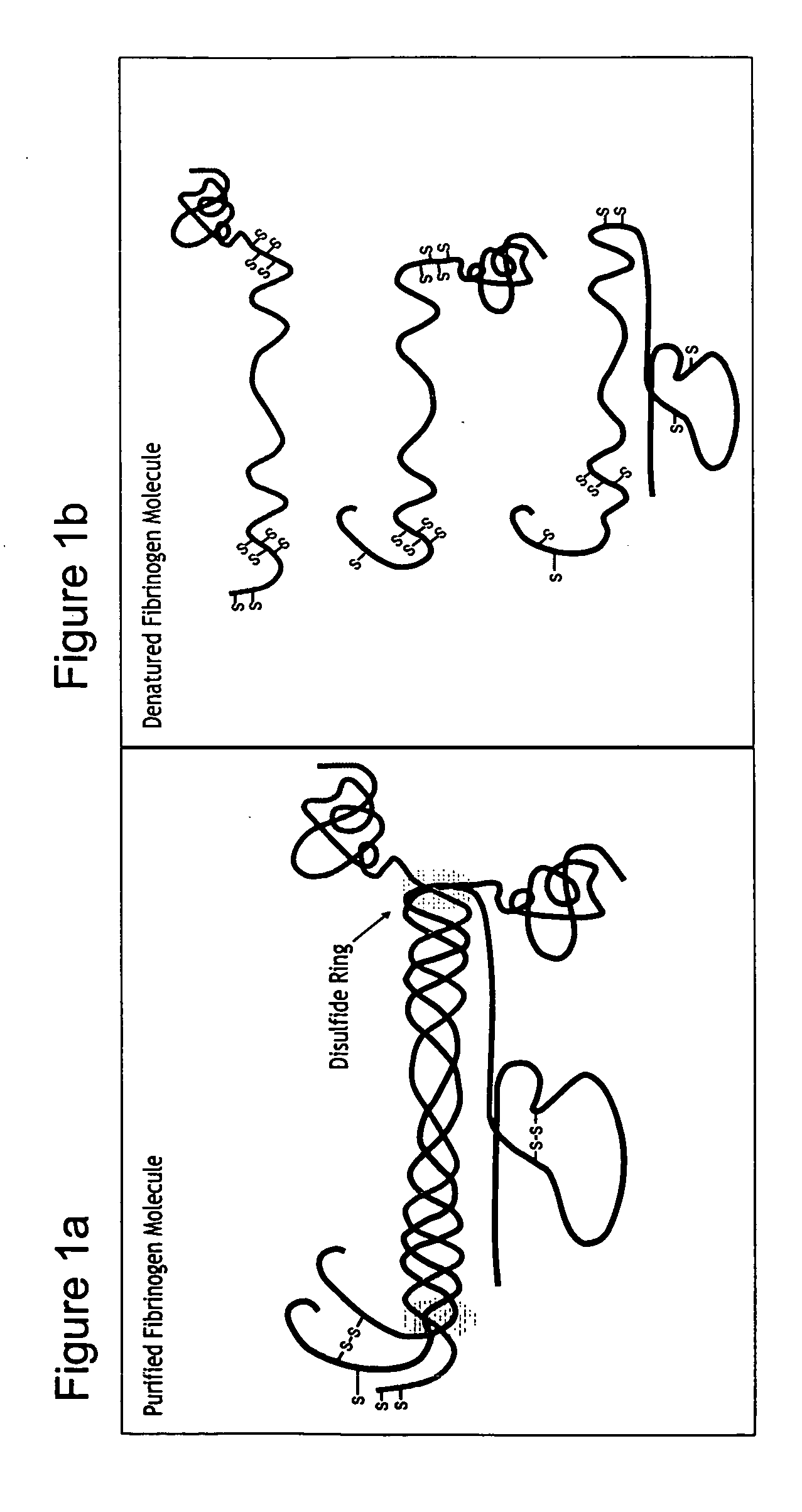
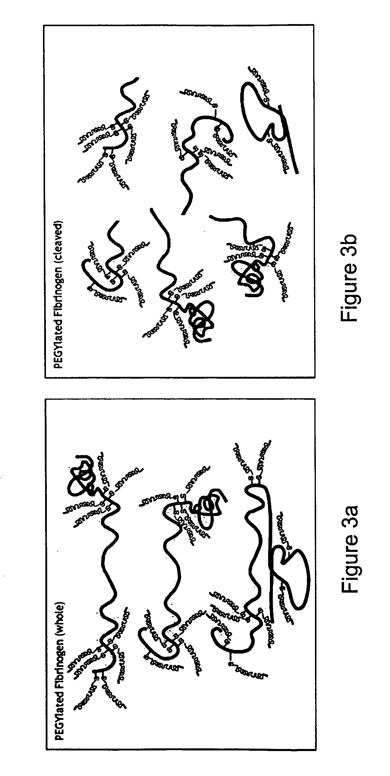
[0150] The blood-borne protein molecule fibrinogen is used to create a hybrid precursor molecule for tissue regeneration [Dikovsky et al., Biomaterials 27(8), 14-96-506, 2006; Almany and Seliktar, Biomaterials, 26(15), 2467-77, 2005]. Fibrinogen is the precursor to the commonly known blood clot protein, fibrin. The fibrinogen molecule contains a set of biological cell-signaling cues specific to cellular remodeling, including the cell-adhesive sequence Arg-Gly-Asp (RGD) and a protease degradation substrate [Herrick, Int J Biochem Cell Biol, 31(7) 741-6,1999]. The fibrinogen molecules are transformed into a biosynthetic hybrid precursor using four sequential steps: 1) fibrinogen denaturation; 2) cleavage (optional); 3) PEGylation; 4) and 3-D matrix formation (optional). Each of these steps is detailed infra.

[0151] Step 1—Fibrinogen Denaturation: Denatured fibrinogen was prepared by incubating purified fibrinogen in a reducing a solution containing a high excess ...

example 2

Denatured PEGylated Fibrinogen Degradation

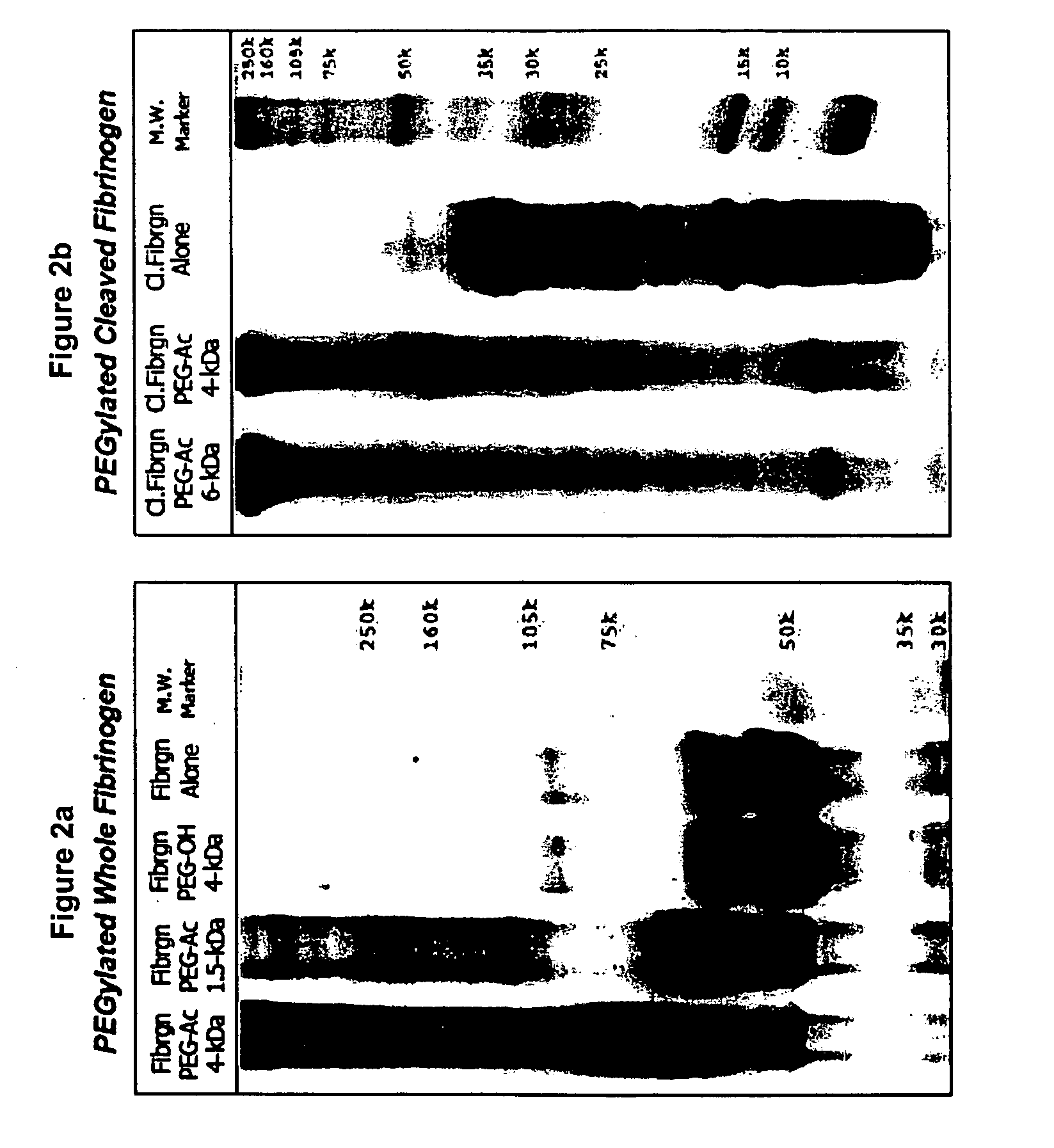
[0157] The degradation of denatured or fragmented PEGylated fibrinogen into PEGylated fibrinogen degradation products was verified experimentally using an in vitro degradation assay. Basically, PEGylated denatured / fragmented fibrinogen fragments are formed into a matrix to recreate a 3-D matrix which can be implanted into a site-specific defect.

[0158] Several variations of the PEGylated fibrinogen fragments are used in these experiments, including cleaved and whole protein with either 6-kDa or 10-kDa PEG-DA attached to the protein. Protease degradation of the denatured PEGylated fibrinogen is measured by colorimetrically assessing the release of the PEGylated fibrinogen degradation products into the supernatant during the course of the experiments. Cylindrical plugs are cast in 5 mm diameter silicon tubes from 100 μl aliquots of PEG-fibrinogen precursor solution containing photoinitiator. The degradation is quantified by labeling the PEGyl...

example 3

Tissue Culture Assay

[0165] The protease-mediated release of the denatured PEGylated fibrinogen products from intact 3-D materials may have a profound chemotactic effect on in vitro tissue cultures of various types. It is assumed that when released from the 3-D matrix, the denatured PEGylated fibrinogen degradation products may facilitate the chemotactic invasion of cells, including vascular smooth muscle cells, chondrocytes, and embryonic stem cells, into the 3-D matrix containing the bound PEGylated fibrinogen. Therefore, the chemotactic potential of the denatured PEGylated fibrinogen degradation products was assayed by an in vitro tissue culture assay designed in part for this task.

[0166] Chemotaxis assay—A cell invasion assay was employed using smooth muscle tissue encapsulated by a PEGylated fibrinogen matrix (10 kDa PEG). Dense tissue constructs composed of smooth muscle cell-seeded collagen gels placed inside PEG-fibrinogen hydrogels. The smooth muscle tissue constructs were...


PUM

PropertyMeasurementUnit
PEG-DAaaaaaaaaaa
PEG-DAaaaaaaaaaa
molar ratioaaaaaaaaaa

Abstract

A method of treating a disorder characterized by tissue damage is provided. The method comprising providing to a subject in need-thereof a composition which comprises a synthetic polymer attached to denatured fibrinogen or a therapeutic portion of the fibrinogen, the composition being formulated for releasing the therapeutic portion of the fibrinogen in a pharmacokinetically regulated manner, thereby treating the disorder characterized by tissue damage or malformation.

Description

RELATED APPLICATIONS [0001] This is a continuation-in-part of PCT Patent Application No. PCT / IL2004 / 001136 filed Dec. 15, 2004, which claims the benefit of U.S. Provisional Patent Application No. 60 / 530,917 filed Dec. 22, 2003. The contents of the above applications are all incorporated by reference.FIELD AND BACKGROUND OF THE INVENTION [0002] The present invention relates to compositions and methods for treating disorders associated with tissue damage, loss, or malformation. [0003] Tissue engineering, i.e., the generation of new living tissues in vitro, is widely used to replace diseased, traumatized or other unhealthy tissues. The classic tissue engineering approach utilizes living cells and a basic scaffold for cell culture (Langer and Vacanti, 1993; Nerem and Seliktar, 2001). Thus, the scaffold structure attempts to mimic the natural structure of the tissue it is replacing and to provide a temporary functional support for the cells (Griffith L G, 2002). [0004] Tissue engineering...

Claims


Application Information

Patent Timeline
Patent Type & Authority Applications(United States)
IPC IPC(8): A61K38/38A61F2/28A61K47/48A61L24/10A61L27/22A61L27/38C07K14/75C12N5/00
CPCA61K47/48215A61K38/00A61K47/48784A61L24/106A61L27/18A61L27/225A61L27/227A61L27/24A61L27/38A61L27/52C07K14/75C12N5/0068C12N2533/30C12N2533/40C12N2533/50C12N2533/52C12N2533/54A61K47/48292C08L71/02A61K47/60A61K47/6435A61K47/6903A61K38/363A61P17/02A61P19/00A61P43/00A61K9/0024A61K9/06A61K35/34
Inventor SELIKTAR, DRORALMANY, LIORA
Owner REGENTIS BIOMATERIALS
Why Eureka
Try Eureka