
Porous beta-tricalcium phosphate granules for regeneration of bone tissue
a beta-tricalcium phosphate and bone tissue technology, applied in the direction of prosthesis, drug composition, organic carrier, etc., can solve the problems of excessive resorption rate, brittle porous ceramics, and inability to be easily shaped by the practitioner, so as to improve the regeneration of bone tissue
- Summary
- Abstract
- Description
- Claims
- Application Information
AI Technical Summary
Benefits of technology
Problems solved by technology
Method used
Image

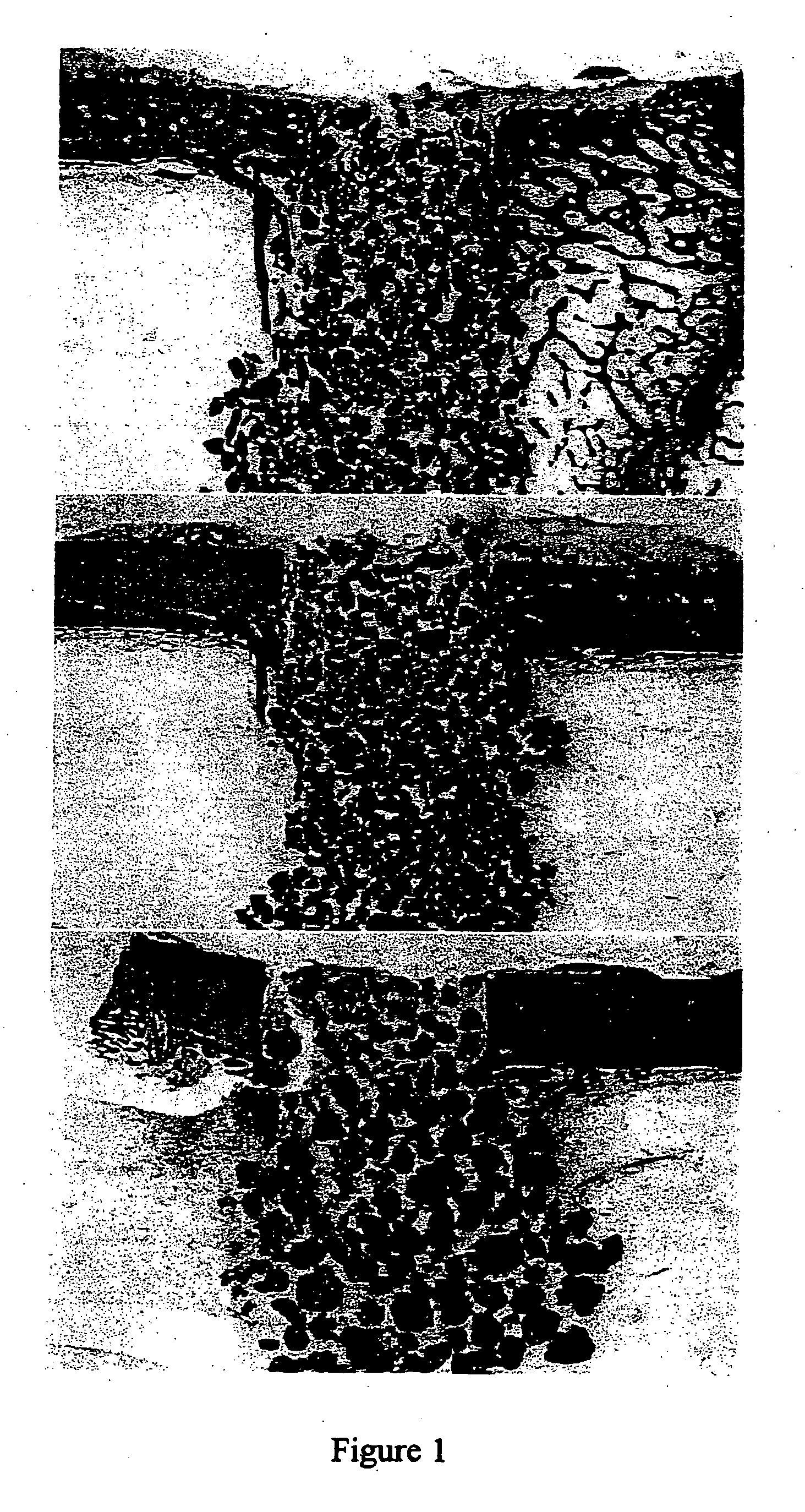
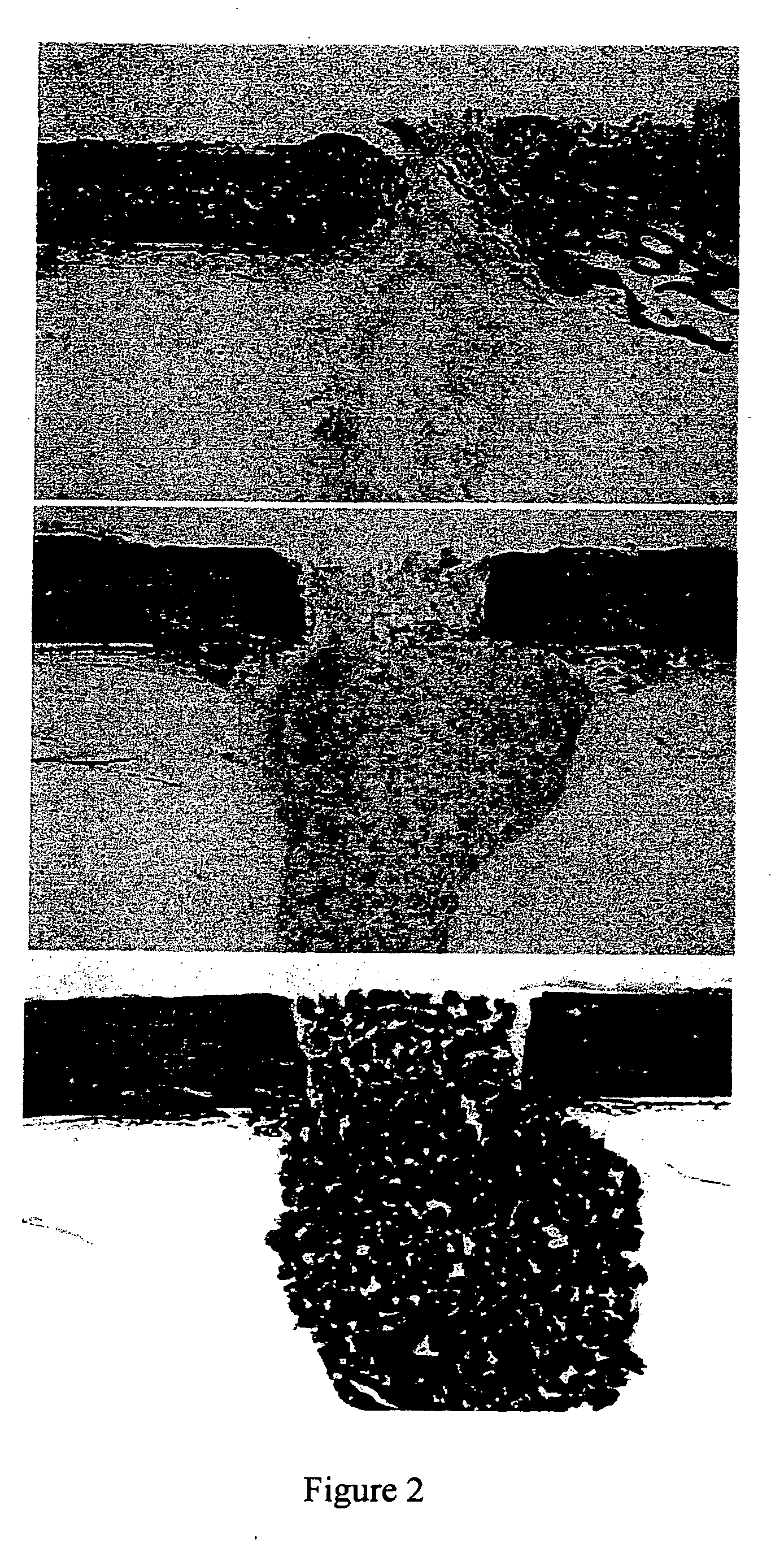
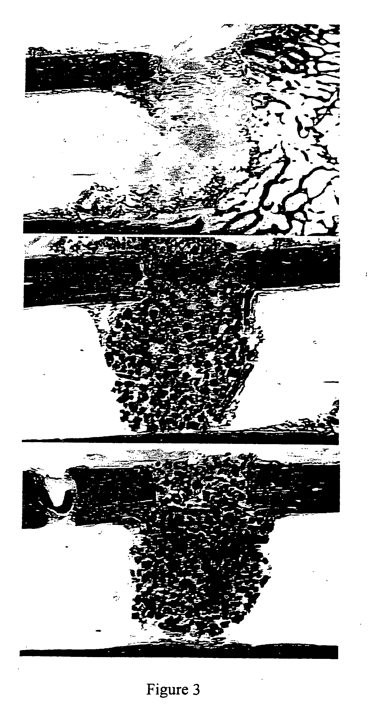
Examples
example 1
Preparation of Tricalcium Phosphate
[0156] A slurry of lime (calcium oxide-hydroxide) is prepared and dilute phosphoric acid is added dropwise to the slurry, which is stirred constantly. The molar proportion of calcium oxide to phosphoric acid is 3:2. The product characteristics are evaluated by X-ray diffraction and adjustments are made to the proportions if required. The resultant slurry is harvested by spray drying. If the slurry is harvested by filtration, the dried cake is crushed to a fine powder of amorphous TCP. The particle size of the amorphous TCP is preferably smaller than 10 μm.
example 2
Preparation of β-TCP Granule
[0157] The TCP powder was mixed with polystyrene beads (NUNC A / S-Denmark)(0-160 μm beads). The 10% polyvinyl pyrrolidone (PVP) granulating solution was prepared by adding PVP C-30 (Plasdone C-30, ISP technologies lot # TX 60810) in small portions in a beaker or flask of stirring water until the solution was clear. About 37 ml of 10% PVP solution was added to the TCP mixture in 5 ml increments to form a crumbly mass. As illustrated in Table 1, mixtures were prepared with different proportions of pore-forming beads and TCP.
TABLE 1bead composition(w / w)beads (g)TCP (g)12.5%12.587.5 25%12.537.537.5%18.7531.25 50%23.7523.75
[0158] The crumbly mass was passed through <500 μm, 500-1000 μm, or 1000-1700 μm sieves under a vibrating motion to produce wet granules having the corresponding particle size ranges. The sieved material was dried under vacuum at 105° C. for 2-3 hours.
[0159] The dried granules then underwent a burn off cycle to vaporize / carbonize the po...
example 3
Rat Model Bioassay for Bone Induction
[0161] This assay consists of implanting samples in subcutaneous sites in recipient rats under ether anesthesia. Male Long-Evans rats, aged 28-32 days, may be used. A vertical incision (1 cm) is made under sterile conditions in the skin over the thoracic region, and a pocket is prepared by blunt dissection. Approximately 25 mg of the test sample is implanted deep into the pocket and the incision is closed with a metallic skin clip. The day of implantation is designated as day one of the experiment. Implants are removed at varying times thereafter (i.e. 12 days, 18 days). The heterotrophic site allows for the study of bone induction without the possible ambiguities resulting from the use of orthotropic sites.
[0162] Bone growth is determined biochemically by calcium content of the implant. Calcium content, is proportional to the amount of bone formed in the implant. Bone formation therefore is calculated by determining the calcium content of the ...
PUM
 Abstract
Description
Claims
Application Information