
Methods and systems for sampling, screening, and diagnosis

a sampling and screening technology, applied in the field of methods and systems for sampling, screening and diagnosis, can solve the problems of difficult and time-consuming isolation and identification of unknown microorganisms, difficult to predict which microorganisms will be most useful at a given site, and cannot always be easily applied to real-world situations, etc., to achieve the effect of being reusable or disposable, and being simple to mak

Inactive Publication Date: 2007-07-12
THE JOHN HOPKINS UNIV SCHOOL OF MEDICINE
View PDF9 Cites 48 Cited by
  • Summary
  • Abstract
  • Description
  • Claims
  • Application Information

AI Technical Summary

Benefits of technology

[0009] The present invention provides improved devices, systems, and methods for monitoring environments, e.g., for use in bioremediation, bioprospecting or medical testing and screening. The devices of the invention can be simply made, can be reusable or disposable, and can be used without contaminating the environment being monitored.
[0010] The devices can also be made to be compatible with standard automated sample handling systems, thereby permitting automated, high-throughput analysis of samples obtained from the environment under study. Analysis of microorganism-containing samples can be performed using mass spectrometric methods, which can provide rapid, accurate determination of microbial species and / or determine the levels or types of proteins or other cell products produced in a given environment.
[0011] The inventive device is useful for monitoring sensitive environments in which contamination is to be avoided. In certain embodiment, the device includes an effluent collection reservoir, for collecting the effluent from the capillary compartments or reagent reservoirs. In this way, release of materials from the interior of the device (fluids, biological organisms or cells, chemical compounds, test compounds, and the like) can be avoided. Thus, in one embodiment, the invention provides an improved, lower cost method for environmental monitoring and bioprospecting.
[0013] In another embodiment, the invention provides an improved bioremediation assessment method and tool that will enable the automated, large-volume, high-throughput analysis of bioremediation sites.
[0017] In another aspect, the invention provides a method for rapidly determining the optimal growth conditions for a cell in an environment.

Problems solved by technology

Methods for detecting the growth of microorganisms are well-developed in microbiology laboratories; however, such techniques cannot always be easily applied to real-world situations.
For example, isolation and identification of an unknown microorganism can be difficult and time-consuming, particularly when the microorganism is not suited to growth under typical laboratory conditions.
While bioremediation has been demonstrated to work in some cases, it is often difficult to predict which microorganisms will be most useful at a given site, or which conditions will most effectively promote the bioremediation process.
Without considerable information about the types of pollutants and indigenous microbes present at a contaminated site, the selection of appropriate bioremediation conditions is difficult.
Unfortunately, due to the relatively inaccessible nature of some contaminated sites (for example, underground water), obtaining such information can be difficult.
Conventional sampling and testing can be labor-intensive and time consuming.
The inability to easily and rapidly obtain accurate sampling information also hinders efforts to discover novel microorganisms or natural products, a process often referred to as bioprospecting.
However, without adequate means for obtaining complete information about the prevailing conditions and microbial communities in these areas, bioprospecting efforts can be slow and difficult.
While many approaches to these problems have been proposed, few methods have been developed which are rapid, inexpensive, and easily customized for the study of a wide variety of microorganism types and environments.
However, the device cannot simultaneously collect multiple samples.
While many approaches to this problem have been suggested, few methods are capable of detecting species of interest without extensive sample clean-up and purification steps, which can be time-consuming and expensive.

Method used


Image

  • Methods and systems for sampling, screening, and diagnosis
  • Methods and systems for sampling, screening, and diagnosis
  • Methods and systems for sampling, screening, and diagnosis

Examples


example 1

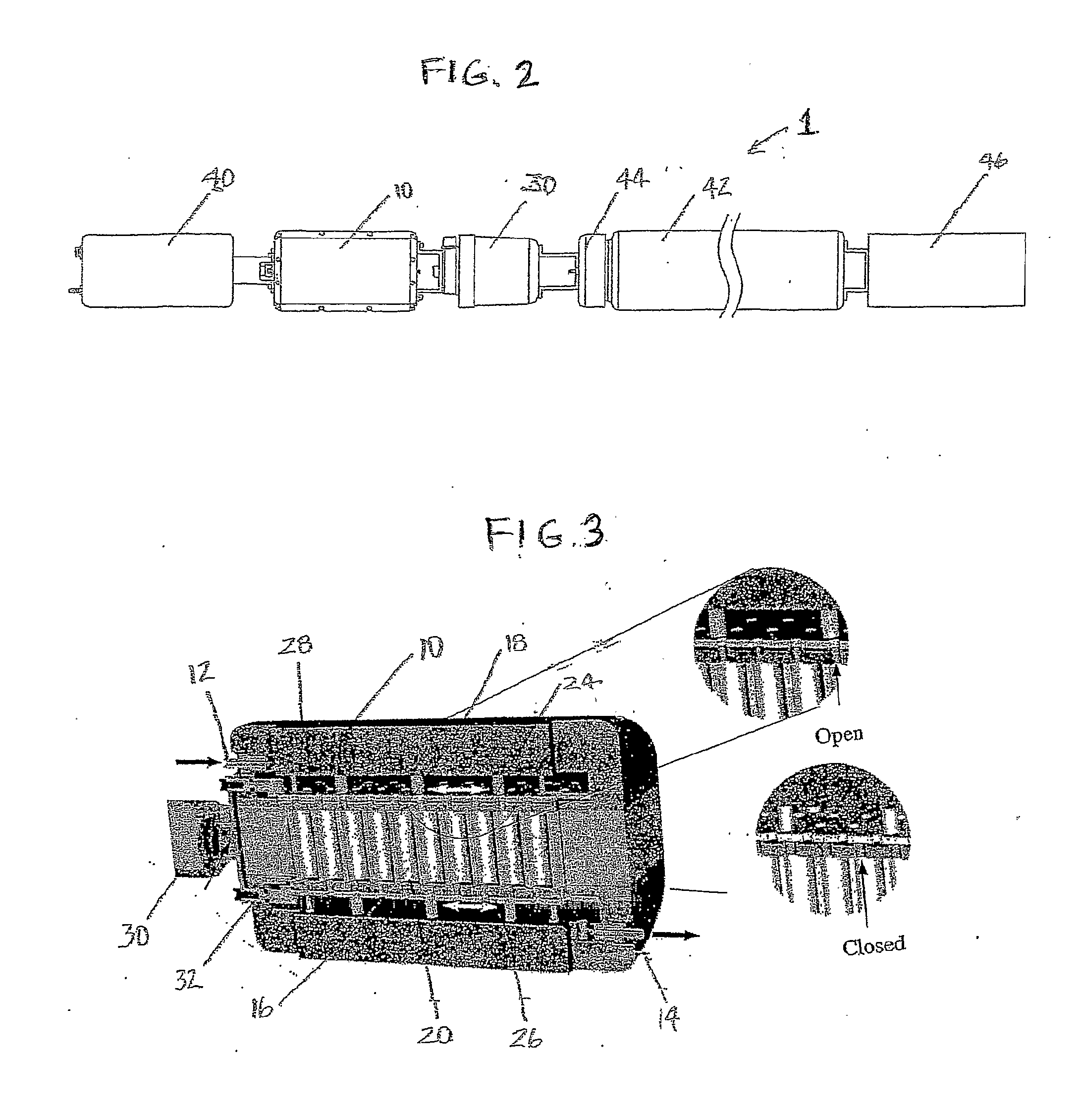
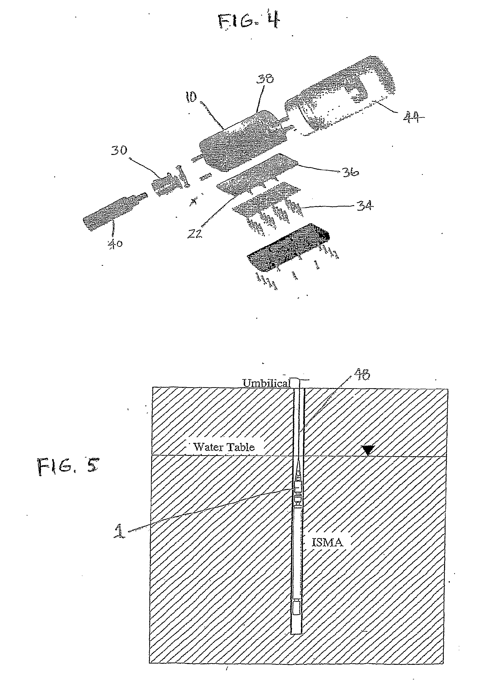
[0116] One embodiment of the present invention takes the form of an in situ microcosm array (ISMA) sampler or testing device 1. This embodiment of the device is well-suited for analysis of environments such as sub-surface water sources or aquifers. As shown in FIGS. 1-4, its principal components include: a housing or container 10 having a fluid inlet 12 and outlet 14, a plurality of capillary microcosms 16 situated within this housing, with these capillaries 16 making up what is referred to as a microcosm array, each of these capillaries 16 having an inlet 18 and outlet 20 that are configured so as to allow for fluid flow through the capillaries 16, each of these capillaries contains an optional filtration material 22 that is selected for its ability to foster microorganism collection in the individual capillaries, upper 24 and lower 26 valve plates having openings 28 that are configured to be alignable with the capillary inlets 18 and outlets 20, a pneumatic cylinder 30 with coupli...

example 2

[0121] In this Example, cells of a bacterium (S. wittichii strain RW1) were identified using MALDI TOF mass spectrometry and multidimensional mass spectrometry in conjunction with peptide mass fingerprinting and peptide sequencing.

[0122] Culturing of Strain RW1. Liquid cultures of S. wittichii strain RW1 (DSMZ 6014) were grown at 30° C. in a water bath shaker in M9 phosphate-buffered minimal medium supplemented with (i) dibenzofuran (DF) crystals (Sigma-Aldrich; Milwaukee, Wis.), (ii) 50 mM glucose, or (iii) both. Saturated DF medium contained approximately 3-5 mg 1−1 of the binuclear aromatic compound in the dissolved phase. Turbidity of the cultures was monitored using a DR / 4000U spectrophotometer (Hach, Loveland, Colo.) at a wavelength of 560 nm. Viable bacteria were enumerated by plate counts using M9 medium supplemented with 1.5% agar (Difco, Franklin Lakes, N.J.) and 5 mM sodium benzoate. Negative control samples composed of cells of RW1 lacking the dioxin dioxygenase were ob...

example 3

[0146] In this Example, a parasite is detected by use of MALDI TOF MS.

[0147] Schistosomes are parasites in a wide variety of warm-blooded hosts. In mammals and humans, they cause serious diseases. It is estimated that hundreds of millions of people worldwide are infected with schistosomes. Infection occurs when cercaria, a free-swimming larval form of the parasite found in contaminated water, penetrate the skin of the mammalian host.

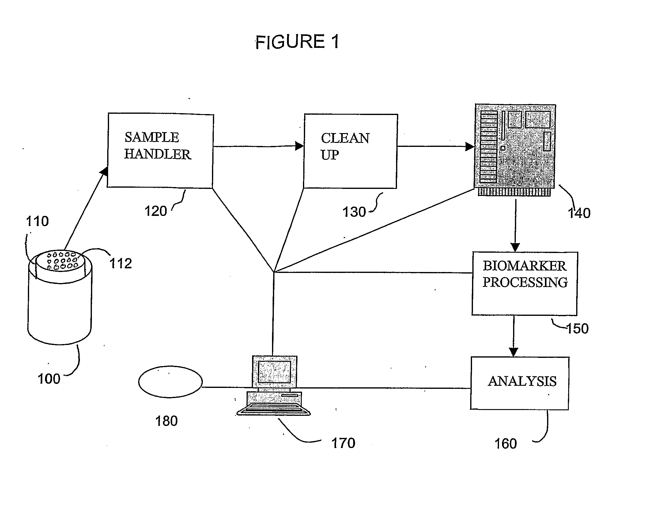
[0148] Detection of the cercariae in water is conventionally performed by skimming the cercariae from the surface of the water, followed by visual observation by a parasitologist. As an alternative, use of MALDI TOF MS was investigated. A schematic representation of the detection method is shown in FIG. 7 (some optional steps are also shown in FIG. 7).

[0149] Methods. Lyophilized cercariae of Schistosoma mansoni were divided into fractions of approximately 2,500 cercariae and resuspended in 50 mM ammonium bicarbonate. Cells were disrupted using a Fishe...


PUM


Abstract

Apparatus, systems, and methods for detecting, screening and sampling of cells are disclosed.

Description

CROSS-REFERENCE TO RELATED APPLICATIONS [0001] This application claims the benefit of U.S. Provisional Application Ser. Nos. 60 / 541,781, filed Feb. 4, 2004; 60 / 577,790, filed Jun. 8, 2004; and 60 / 619,621, filed Oct. 18, 2004. The contents of each of these applications is hereby incorporated herein by reference.BACKGROUND OF THE INVENTION [0002] Biological monitoring of the growth of cells has applications in fields such as microbiology and medicine. Methods for detecting the growth of microorganisms are well-developed in microbiology laboratories; however, such techniques cannot always be easily applied to real-world situations. For example, isolation and identification of an unknown microorganism can be difficult and time-consuming, particularly when the microorganism is not suited to growth under typical laboratory conditions. [0003] In some cases it is important to study a microorganism in its natural or adopted environment. For example, bioremediation is a recent approach to dec...

Claims


Application Information

Patent Timeline
IPC IPC(8): C12Q1/04B01L3/00C12M1/26C12M1/34C12Q1/24C12Q1/26G01N1/00G01N33/68
CPCB01L3/5025H01J49/04B01L2300/0829B01L2300/0838C12Q1/04C12Q1/24C12Q1/26G01N1/10G01N33/6818G01N33/6848G01N2333/90241C12Q1/70G01N33/5008G01N2500/00G01N2500/10B01L2200/0668
Inventor HALDEN, ROLF U.
Owner THE JOHN HOPKINS UNIV SCHOOL OF MEDICINE
Learn More