
Methods and apparatuses for growing cells

a cell culture and cell technology, applied in the field of cell culture, can solve the problems of limited surface area, difficult scaling up, and inability to develop an economical method for mass production of es and eg cells, and achieve the effect of increasing the perfusion flow rate over the stem cells

Inactive Publication Date: 2007-08-02
THE OHIO STATE UNIV RES FOUND
View PDF6 Cites 18 Cited by
  • Summary
  • Abstract
  • Description
  • Claims
  • Application Information

AI Technical Summary

Benefits of technology

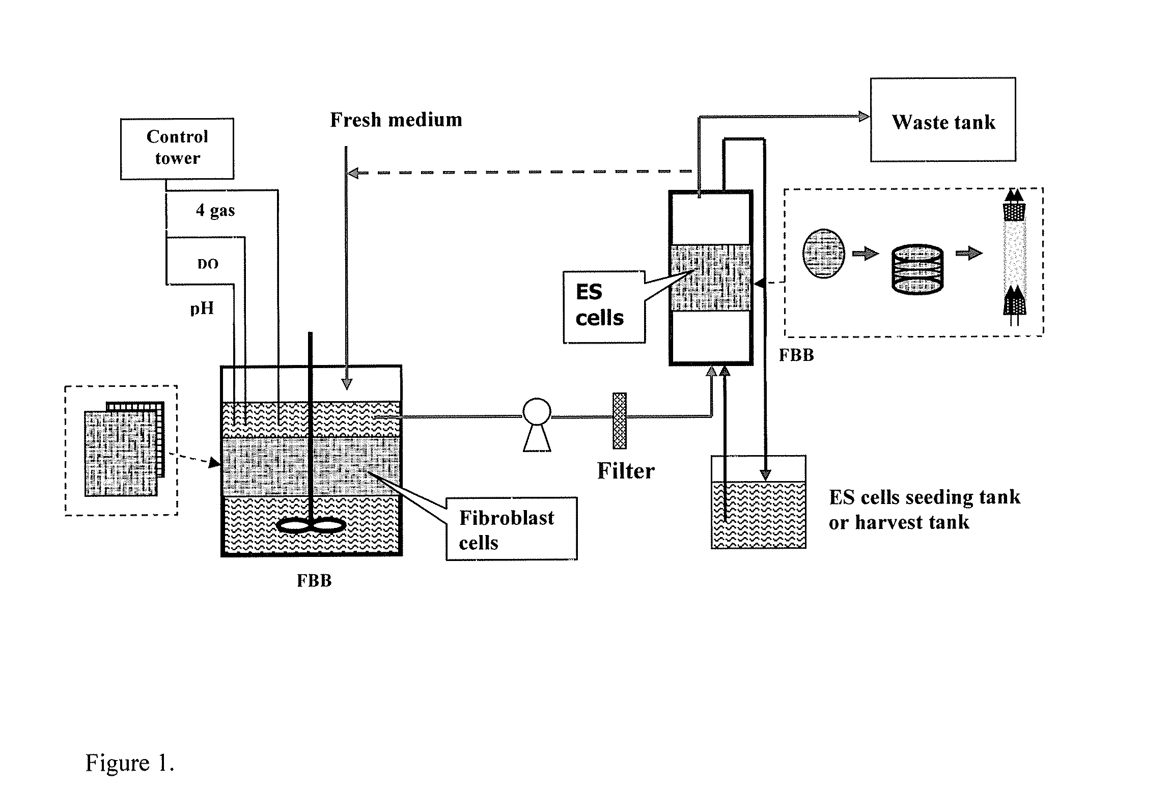
[0010] The various embodiments allow feeder cells to be separated from the stem cells, so as not to contaminate the final stem cell preparation. This physical separation also eliminates the needs of expensive cytokines and growth factors that are required for conventional methods of culturing stem cells.
[0015] The methods and apparatuses further provide methods of culturing stem cells comprising: growing fibroblast feeder cells on a three-dimensional scaffold; perfusing the fibroblast cells with a cell culture medium to form fibroblast cell-conditioned cell culture medium; and growing the stem cells on a three-dimensional scaffold perfused with the fibroblast cell-conditioned cell culture medium. Some embodiments comprise filtering the fibroblast cell-conditioned medium before perfusing the stem cells with the medium. The stem cells may be harvested by steps that include contacting the stem cells with an enzyme chosen from accutase, trypsin and collagenase and / or increasing the perfusion flow rate over the stem cells.

Problems solved by technology

Although the demand for ES and EG cells is high and expected to grow rapidly once their biomedical applications have been established, there has been little effort aimed at developing an economical method for mass production of ES and EG cells.
Currently, the expansion of ES cells is based on common laboratory procedures carried out in two-dimensional (2-D) cell culture systems such as T-flasks, which are limited by the available surface area and difficult to scale up.
Furthermore, the culture surface needs to be pre-coated with expensive extracellular matrix proteins, such as gelatin for murine (mES) and Matrigel for human (hES) ES cells.
These expensive, labor intensive, and time consuming 2-D culturing methods cannot meet the projected market demand for ES cells.
Current supplies of ES and EG cells are limited by the available cell sources, and there is a need to develop a scalable method for mass production of stem cells for biomedical applications.
Current two-dimensional (2-D) culture systems are limited by space and low specific surface area available for cell adhesion and require coating the culture surfaces with expensive extracellular matrix proteins such as gelatin for mouse (mES) and Matrigel for human embryonic stem (hES) cells.
Using the feeder layer or the co-culture method generates a complication in their application due to the cell source contamination from the feeder layer cells when harvesting the ES cells.
However, these growth factors are expensive and their use could impede the scale up of the ES cell production process.

Method used


Image

  • Methods and apparatuses for growing cells
  • Methods and apparatuses for growing cells
  • Methods and apparatuses for growing cells

Examples


example 1

Two-Stage 3-D Bioreactor for Stem Cell Expansion

[0049] Materials and Methods

[0050] Cultures and Media

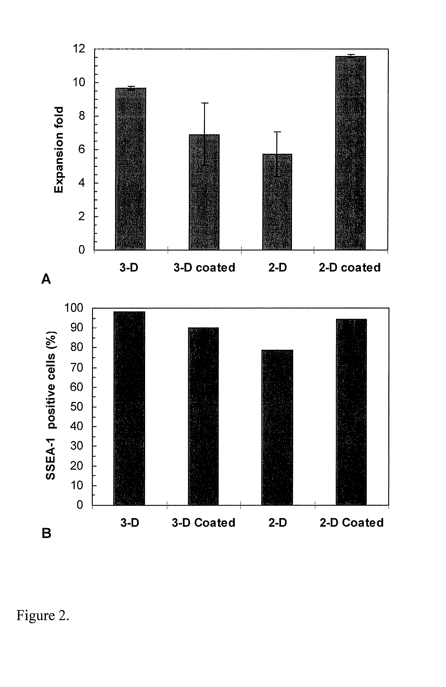
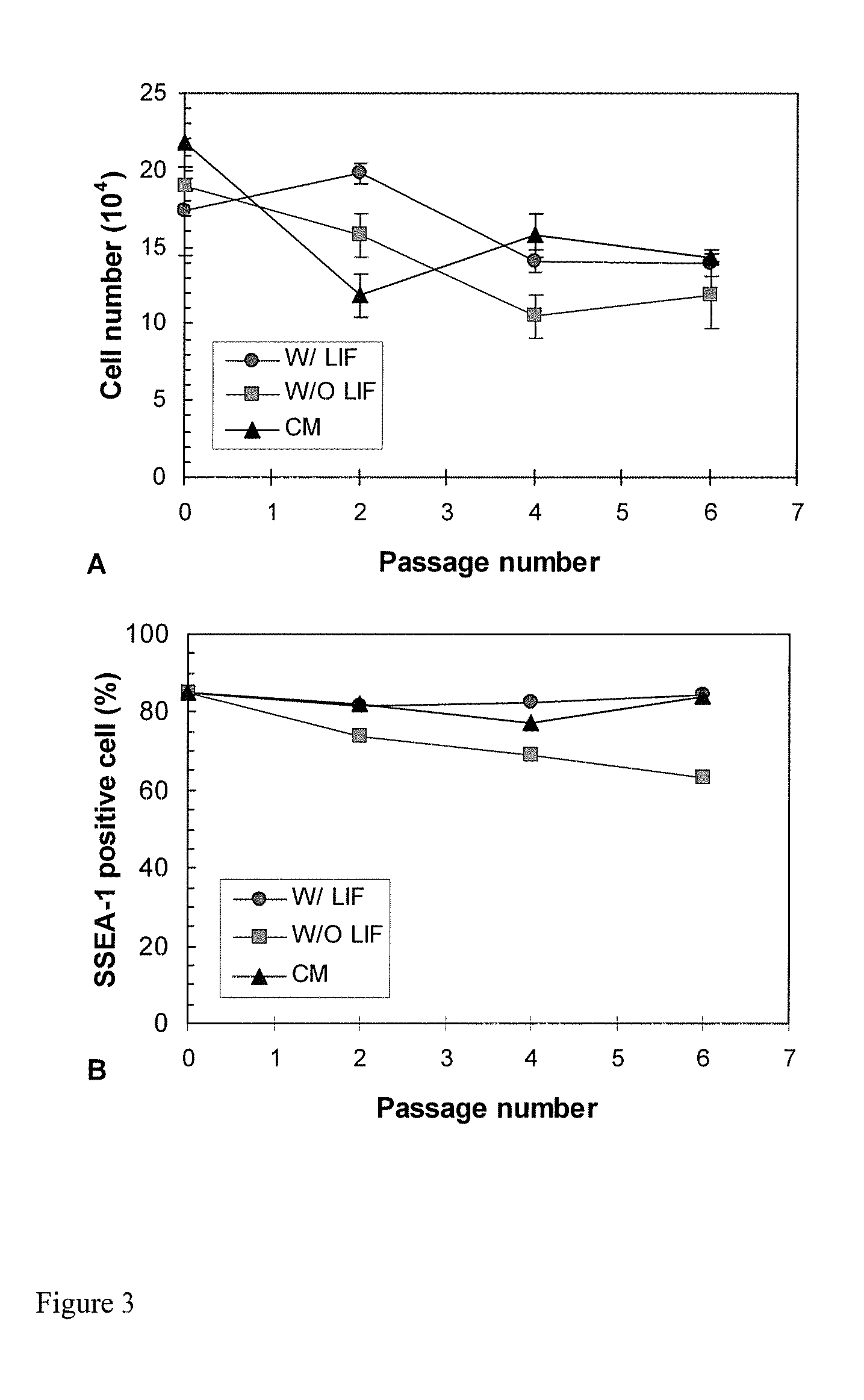
[0051] Murine ES D3 (mES) cells (CRL-1934, ATCC) were maintained on gelatin pre-coated T-flasks containing the ES growth medium, which consisted of the knock-out Dulbecco's Modified Eagle's Medium (DMEM) with 10% fetal bovine serum (FBS), 50 U / ml penicillin, 50 μg / ml streptomycin, 0.1 mM non-essential amino acids, 2 mM L-glutamine, 100 μM β-mercaptoethanol (Sigma, St. Louis, Mich.) and 100 μM leukemia inhibitory factor (LIF) (Chemicon, Temecula, Calif.). Both STO (CRL-1503, ATCC) and mouse embryonic fibroblast (MEF) (SCRC-1045, ATCC) cells were cultured in DMEM with 10% FBS, and they were used to prepare the conditioned media described later. Human ES (hES) cells (SCRC-2002, ATCC) were maintained on Matrigel (BD, San Jose, Calif.) coated T-flasks and cultured in the MEF conditioned medium described below supplemented with 4 ng / ml bFGF. Unless otherwise noted, these T-flask culture...

example 2

Neural Differentiation from Embryonic Stem Cells in Different Culture Systems

[0132] Materials and Methods

[0133] Cultures and Media

[0134] ES D3 cells (CRL-1934) obtained from ATCC were maintained on gelatin pre-coated T-flasks consisting knock-out Dulbecco's Modified Eagle's Medium (DMEM) supplemented with 10% fetal bovine serum (FBS), 50 U / mL penicillin, 50 μg / mL streptomycin, 0.1 mM non-essential amino acids, 2 mM L-glutamine, 100 μM β-mercaptoethanol (Sigma) and 100 μM leukemia inhibitory factor (LIF) (Chemicon). For neural differentiation, LIF was excluded in the above ES medium and either retinoic acid (10−7 M) or an astrocyte-conditioned medium (30% v / v) was added to induce neural differentiation.

[0135] Astrocytes (CRL-2253) from ATCC were cultured in DMEM with 10% FBS. Approximately three to five million astrocyte cells were inoculated to a PET matrix submerged in 110 mL of DMEM with 10% FBS in a 250-mL spinner flask. The medium was refreshed at day six and cells were cult...


PUM


Abstract

Methods of culturing stem cells including growing fibroblast cells on a three-dimensional scaffold, perfusing the fibroblast cells with a cell culture medium to form fibroblast cell-conditioned cell culture medium, and growing the stem cells on a three-dimensional scaffold perfused with the fibroblast cell-conditioned cell culture medium are presented. Multi-stage bioreactors for growing stem cells, comprising a first fibrous bed bioreactor in fluid communication with a second fibrous bed bioreactor are also presented.

Description

CROSS REFERENCE TO RELATED APPLICATIONS [0001] This application claims priority to and any other benefit of U.S. Provisional Application Ser. No. 60 / 734,879, filed on Nov. 9, 2005, the entire content of which is incorporated by reference herein.DESCRIPTION OF THE INVENTION [0002] 1. Field of the Invention [0003] The methods and apparatuses generally relate to culturing of cells, particularly stem cells. [0004] 2. Background [0005] Embryonic stem (ES) cells derived from the inner cell mass of blastocyst have unlimited proliferation potential and are totipotent. Embryonic germ (EG) cells originate from the reproductive cells of fetal cadaver tissue in the gonad ridge. EG cells are further along in development, and therefore cannot be derived from embryos, but instead must be isolated from fetal tissue. EG cells have the ability to form all three germ layers and therefore potentially all the organs of the body. Therefore, both are ideal cell sources for tissue engineering and cell ther...

Claims


Application Information

Patent Timeline
IPC IPC(8): C12N5/06C12M3/00
CPCC12M23/58C12M29/10C12M25/14C12M35/08C12M29/04
Inventor YANG, SHANG-TIANOUYANG, ANIL
Owner THE OHIO STATE UNIV RES FOUND
Learn More