Prenatal Diagnosis Using Cell-Free Fetal DNA in Amniotic Fluid

a fetal dna and amniotic fluid technology, applied in the field of prenatal diagnosis using cell-free fetal dna in amniotic fluid, can solve the problems of enormous medical care costs, numerical and structural chromosomal abnormalities over the whole genome, tedious, time-consuming and laborious, etc., and achieve the effect of faster determination of the “molecular karyotype” of the fetus

Inactive Publication Date: 2007-09-13
BIANCHI DIANA W +2
View PDF99 Cites 190 Cited by
  • Summary
  • Abstract
  • Description
  • Claims
  • Application Information

AI Technical Summary

Benefits of technology

[0010] The present invention provides an improved system for analyzing a fetus' genetic information. In particular, the present invention for allows the rapid determination of a “molecular karyotype” of the fetus. This molecular karyotype can provide more

Problems solved by technology

Furthermore, genetic disorders and congenital anomalies contribute substantially to long-term disability; they are associated with enormous medical-care costs (A. Czeizel et al., Mutat. Res. 1984, 128: 73-103; Centers of Disease Control, Morb. Mortal. Weekly Rep. 1989, 38: 264-267; S. Kaplan, J. Am. Coll. Cardiol.
Examination of the karyotypes determined by these banding methods can reveal the presence of numerical and structural chromosomal abnormalities over the whole genome.
In addition to tissue sampling and selective staining, conventional banding methods also require cell culturing, which can take between 10 and 15 days depending on the tissue source, and preparation of high quality metaphase spreads, which is tedious, time-consuming and labor-intensive (B. Eiben et al., Am. J. Hum. Genet.
Furthermore, conventional chromosome analysis methods have limited sensitivity, and their standard 450-550 band level of resolution does not allow detection of small or subtle chromosomal aberrations, such as, for example, those associated with microdeletion/microduplication syndromes.
However, in contrast to conventional banding analysis, certain molecular cytogenetic methods

Method used


Image

  • Prenatal Diagnosis Using Cell-Free Fetal DNA in Amniotic Fluid
  • Prenatal Diagnosis Using Cell-Free Fetal DNA in Amniotic Fluid
  • Prenatal Diagnosis Using Cell-Free Fetal DNA in Amniotic Fluid

Examples


example 1

Amniotic Fluid Fetal DNA Isolation and Preliminary Tests

[0251] Frozen amniotic fluid supernatant specimens (38) were obtained from the Tufts-New England Medical Center (Tufts-NEMC) Cytogenetics Laboratory (D. W. Bianchi et al., Clin. Chem. 2001, 47: 1867-1869). All samples were collected for routine indications, such as advanced maternal age, abnormal maternal serum screening results, or detection of a fetal sonographic abnormality. The standard protocol in the Cytogenetics Laboratory is to centrifuge the amniotic fluid sample upon receipt, place the cell pellet into tissue culture, assay an aliquot of the fluid for alpha-fetoprotein and acetyl cholinesterase levels, and store the remainder at −20° C. as a back-up in case of assay failure. After six months, the frozen amniotic fluid supernatant samples are normally discarded.

[0252] The frozen fluid samples obtained from the Cytogenetics Laboratory were initially thawed at 37° C. and then mixed with a vortex for 15 seconds. An aliq...

example 2

Molecular Karyotyping using Cell-Free Fetal DNA from Amniotic Fluid

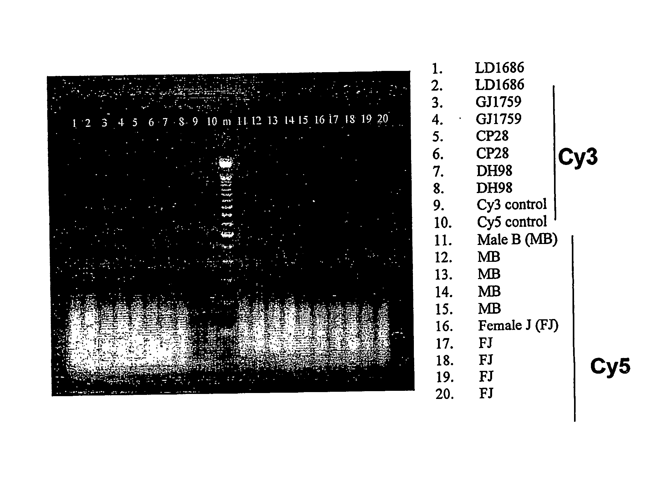
[0257] To determine if cell-free fetal DNA in amniotic fluid could be used for molecular karyotyping, cell-free DNA was extracted from eight frozen amniotic fluid supernatant samples from four known euploid males and four known euploid females. Each sample was ≧10 mL in volume and yielded between 200 and 900 ng of DNA. The samples were sent to Vysis for analysis. The results obtained by Vysis confirmed the quantity of DNA present. The concentration of DNA was adjusted to 25 ng / μL. Samples were labeled with Cy-3™ and Cy-5™ according to the current labeling protocol for the GenoSensor™ Array 300. For each sample, reference male and female DNA of equal quantity was labeled for CGH. After DNase digestion, samples were visualized on a 2% agarose / ethidium bromide gel. As shown in FIG. 1, DNA from samples and controls demonstrated uniform amplification and labeling.

[0258] Samples were combined, added to hybridization buff...

example 3

Use of Amniotic Fluid Cell-Free Fetal DNA in CGH Microarrays to Generate a Molecular Karyotype: Preliminary Studies

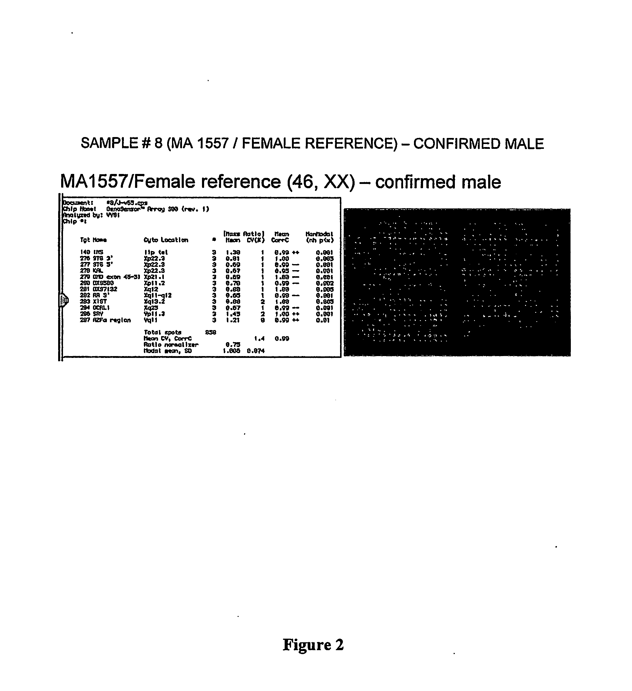
[0262] In a typical analysis, fetal DNA is extracted from stored amniotic fluid supernatant samples with normal and abnormal karyotypes. The samples are then sent to Vysis for analysis. The samples are hybridized to euploid male and euploid female reference DNA on CGH microarrays. The hybridization data is then analyzed and interpreted by the Applicants at Tufts / New England Medical Center.

[0263] Vysis has developed a novel microarray technology system that permits simultaneous assessment of multiple genomic targets. The GenoSensor™ system consists of the following hardware: MacIntosh G3 PowerPC computer with 17″ high resolution display monitor, 1.3 million pixel high-resolution cooled CCD camera, custom-designed optics, automated 6-position filter wheel with 3 filters, and xenon illumination source. The microarray consists of over 1,300 gene loci derived primarily fro...


PUM

PropertyMeasurementUnit
Timeaaaaaaaaaa
Timeaaaaaaaaaa
Ratioaaaaaaaaaa

Abstract

The present invention relates to improved methods of prenatal diagnosis, screening, monitoring and/or testing. The inventive methods include the analysis by array-based hybridization of cell-free fetal DNA isolated from amniotic fluid. In addition to allowing the prenatal diagnosis of a variety of diseases and conditions, and the assessment of fetal characteristics such as fetal sex and chromosomal abnormalities, the new inventive methods provide substantially more information about the fetal genome in less time than it takes to perform a conventional metaphase karyotype analysis. In particular, the enhanced molecular karyotype methods provided by the present invention allow the detection of chromosomal aberrations that are not often detected prenatally such as microdeletions, microduplications and subtelomeric rearrangements.

Description

RELATED APPLICATION [0001] This application claims priority to Provisional Patent Application No. 60 / 515,735, filed Oct. 30, 2003, which is incorporated herein by reference in its entirety.BACKGROUND OF THE INVENTION [0002] Genetic disorders and congenital abnormalities (also called birth defects) occur in about 3 to 5% of all live births (A. Robinson and M. G. Linden, “Clinical Genetic Handbook”, 1993, Blackwell Scientific Publications: Boston, Mass.). Combined, genetic disorders and congenital abnormalities have been estimated to account for up to 30% of pediatric hospital admissions (C. R. Scriver et al., Can. Med. Assoc. J. 1973, 108: 1111-1115; E. W. Ling et al., Am. J. Perinatal. 1991, 8: 164-169) and to be responsible for about half of all childhood deaths in industrialized countries (R. J. Berry et al., Public Health Report, 1987, 102: 171-181; R. A. Hoekelman and I. B. Pless, Pediatrics, 1998, 82: 582-595). In the US, birth defects are the leading cause of infant mortality ...

Claims


Application Information

Patent Timeline
IPC IPC(8): C12Q1/68
CPCC12Q1/6806C12Q1/6883G01N21/6428C12Q2600/156C12Q2565/501C12Q2523/32
Inventor BIANCHI, DIANA W.LARRABEE, PAIGE B.JOHNSON, KIRBY L.
Owner BIANCHI DIANA W
Why Eureka
Try Eureka