Three-dimensional tissue equivalent using macromass culture

a three-dimensional tissue and culture technology, applied in the field of tissue engineering, can solve the problems of insufficient vascular or venous healing, poor healing effect of neuropathic diabetic foot ulcers, and toxic topical antiseptics, such as povidone-iodine, and achieve the effect of improving the healing effect and improving the healing

Inactive Publication Date: 2008-06-19
RELIANCE LIFE SCI PVT
View PDF6 Cites 40 Cited by
  • Summary
  • Abstract
  • Description
  • Claims
  • Application Information

AI Technical Summary

Benefits of technology

[0044]In another embodiment, the dermal fibroblast cells are not lost by leaking through the pores of the porous scaffold or matrix during the culturing step.

Problems solved by technology

Lower extremity skin ulcers can result from arterial or venous insufficiency.
However, randomized controlled clinical trials showed only a 15% acceleration in the healing of neuropathic diabetic foot ulcers.
Topical antiseptics, such as povidone-iodine, are usually considered to be toxic to healing wounds.
Growth factors besides PDGF have not been approved for clinical use and the results of clinical trials have not delivered the expectations generated by preclinical data.
With the advent of tissue engineering, there have been promising results shown by different skin substitutes in efficiently treating chronic wounds, which have otherwise been difficult to heal successfully and often lead to amputation of the limb having the ulcer.
While skin autografts are successful in effecting wound healing, the autografting procedure is invasive, painful and could lead to a secondary non-healing wound.
However the skin substitutes face challenges such as difficult logistics of ordering and using the substitutes due to a cryopreservation requirement, difficulty in maintaining cell viability, poor durability of matrix collagen when exposed to the enzyme-rich wound bed (thereby causing cells to wash away and lose effect), and the thickness of the matrix preventing sufficient diffusion of growth factors from the embedded cells.
Another pertinent problem is the cost of these skin substitutes.
In some tissue equivalents that have been developed, there is significant contraction their original size, which limits such equivalents to specific applications in which the contracted construct can be useful.
However, non-porous supports limit the supply and diffusion of nutrients and gases.
On the other hand, one problem of culturing cells using porous matrices is that a percentage of total cells seeded onto the sponge in the form of cell suspension leak out from the bottom of the sponge onto the base of the culture vessel.
This amounts to loss of cells when seeding, which is an especially critical problem for tissue engineering applications wherein cell sources can be rather limited.
Loss of cells would also result in difficulty in getting reproducible constructs from equally seeded sponges, since variable numbers of cells would remain on the sponges if varying numbers of cells are lost while seeding.

Method used


Image

  • Three-dimensional tissue equivalent using macromass culture
  • Three-dimensional tissue equivalent using macromass culture
  • Three-dimensional tissue equivalent using macromass culture

Examples


example 1

Preparation of the Three Dimensional Tissue Equivalent

I. Cell Isolation and Culture.

[0108]In the present invention, human dermal fibroblasts were isolated from discarded human skin biopsies obtained with written informed consent. The dermis was separated from the epidermis by treatment with Dispase (Sigma, St. Louis, USA). The dermis was minced and digested with 0.01% collagenase in DMEM+10% FCS overnight and then cells were allowed to attach to a culture flask. Cells were cultured in DMEM+10% FCS at 37° C. in 5% CO2 and subcultured using Trypsin-EDTA solution.

II. Preparation of Chitosan Sponges.

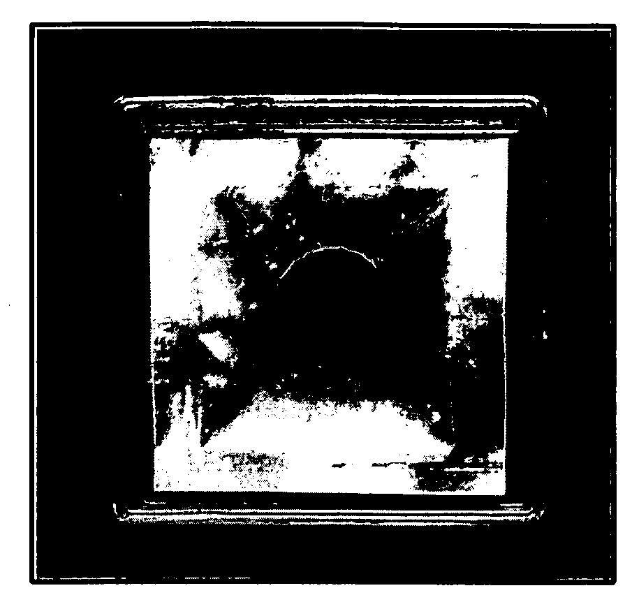
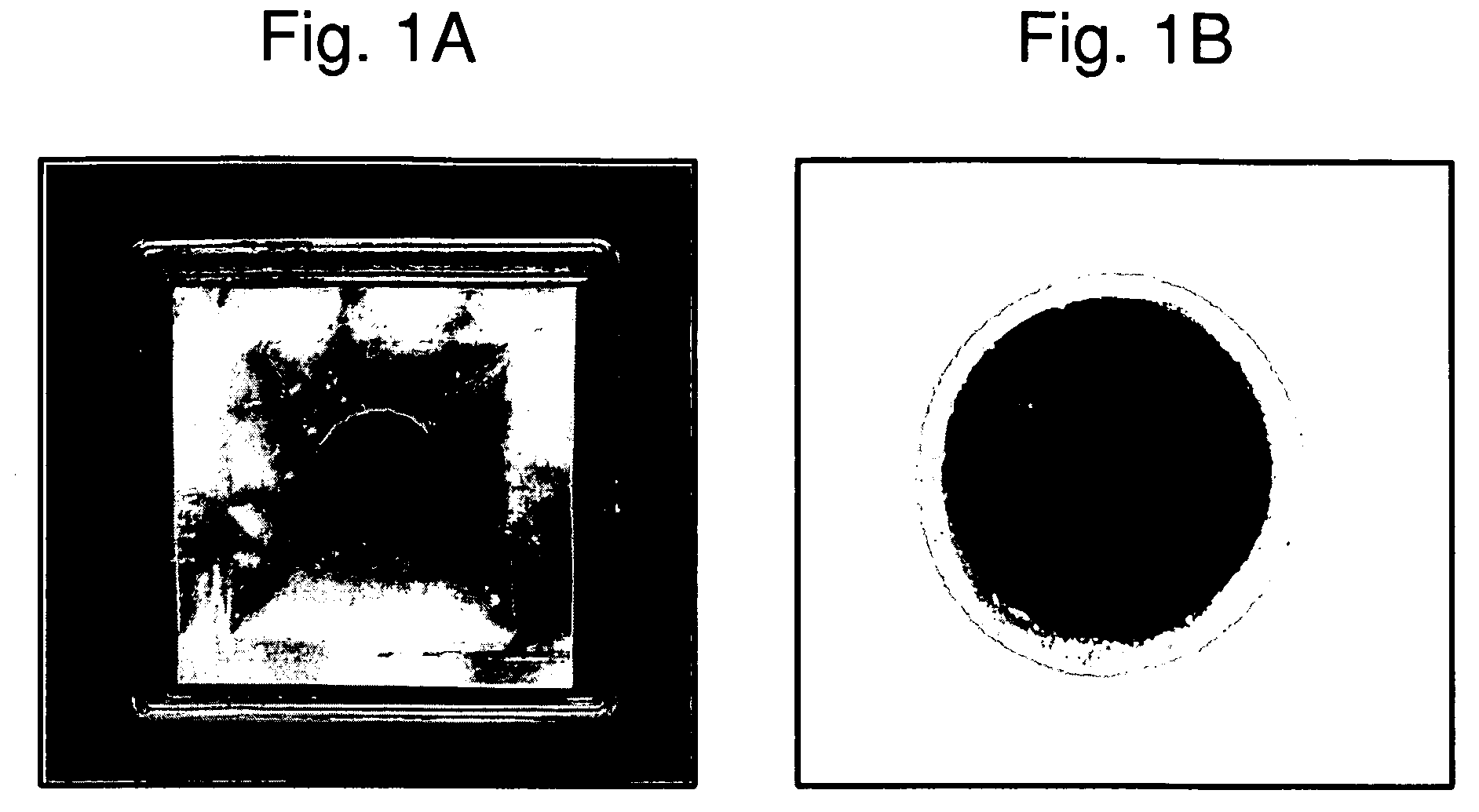
[0109]Chitosan sponges having a diameter of 3.0 cm and thickness of about 1.5 mm were prepared by lyophilization of frozen chitosan solution in 3.5 cm dishes. After lyophilization, the chitosan sponges were stabilized in isopropanol. The chitosan sponges were treated with ammonia and methanol solution. Chitosan sponges were rinsed with water for 3 hours with shaking. Chitosan sponges were th...

example 2

Characterization and Evaluation of Three Dimensional Tissue Equivalents

[0114]The three dimensional tissue equivalent of the present invention was evaluated for safety and effectiveness, and the data are classified below in four categories: (1) Safety, (2) Potency, (3) Purity, and (4) Stability.

1. Safety

A) Sterility

[0115]To ensure that aseptic conditions were maintained from the manufacturing process until final packing, the batches of tissue equivalent of the present invention were tested for sterility to detect the presence of aerobic and anaerobic microbes. This test was performed using the Direct Inoculation method (IP, 1996), which involved inoculating the test sample in two different sterile nutritive media, namely, Fluid Thioglycollate Medium (FTM) and Soybean Casein Digest Medium (SCDM). Absence of growth in the inoculated media during the incubation period of 14 days confirmed the sterility of the samples.

B) Bioburden

[0116]The microbial load in terms of number of colonies ap...

example 3

In Vivo Studies and Toxicology

1. Study of Efficacy and Safety of Tissue Equivalent of the Present Invention in a Wound Healing Animal Model.

[0133]The efficacy and safety of the tissue equivalent of the present invention was assessed by performing a skin wound healing study in severe combined imunodefiicent (SCID) CB17 mice. SCID mice were used since the dermal dressing to be tested consists of human cells, which would undergo xenograft rejection if tested on animals that are non-immuno-compromised. The study was carried out in accordance with CPCSEA guidelines and with IAEC approval. There were four sets of animals, one set for each time period after surgery and application where tissue would be harvested, namely, 4 days, 8 days, 12 days, and 16 days. For each time point, there were three control animals and six animals treated with the tissue equivalent. In the control group, chitosan sponge alone was applied on each wound. In the treated group, the tissue equivalent was applied on...


PUM

PropertyMeasurementUnit
diameteraaaaaaaaaa
thicknessaaaaaaaaaa
thicknessaaaaaaaaaa

Abstract

The present invention provides a three-dimensional tissue equivalent for in-vivo and in-vitro uses. The three dimensional tissue equivalent of the present invention is a non-contractile cellular sheet cultured over a porous scaffold using a macromass culturing technique, for example where the cellular sheet is entirely on one side of a porous sponge. In one embodiment, the present invention provides a dermal wound dressing that comprises a high density cellular sheet of dermal fibroblast cells.

Description

CROSS REFERENCE TO RELATED APPLICATIONS[0001]This application is a continuation-in-part of U.S. patent application Ser. No. 10 / 686,822, filed Oct. 16, 2003, which claims the benefit of India application Ser. No. 912 / MUM / 2002, filed Oct. 18, 2002, now Indian Patent No. 195953, all of which are hereby incorporated by reference in their entirety for all purposes. Related applications, India application Ser. Nos. 2013 / MUM / 2006 and 2014 / MUM / 2006, were also filed in India on Dec. 12, 2006.FIELD OF THE INVENTION[0002]The present invention relates to the field of tissue engineering for in vivo or in vitro uses. The present invention further relates to a non-contractile three-dimensional tissue equivalent as a dermal wound dressing and its methods of preparation. The present invention also relates to therapeutic applications of the non-contractile three-dimensional tissue equivalent and its safety and efficacy evaluation in the treatment of wounds. Alternatively the three-dimensional tissue ...

Claims


Application Information

Patent Timeline
Patent Type & Authority Applications(United States)
IPC IPC(8): A01N63/00C12N5/02C12Q1/20C12N5/00
CPCA61K35/12A61L15/40A61L15/425C12N5/0656A61L27/60G01N33/5011A61L27/3813A61L27/56C12N2533/72
Inventor DESHPANDE, MANISHA SHARADCHANDRARAO, SITHAMRAJU HARINARAYANAWANGIKAR, PRALHAD BALASAHEBKUCHROO, PUSHPA VIKRAM
Owner RELIANCE LIFE SCI PVT
Why Eureka
Try Eureka