
Methods for Implementing Microbeam Radiation Therapy

Inactive Publication Date: 2008-08-14
BROOKHAVEN SCI ASSOCS
View PDF14 Cites 96 Cited by
  • Summary
  • Abstract
  • Description
  • Claims
  • Application Information

AI Technical Summary

Benefits of technology

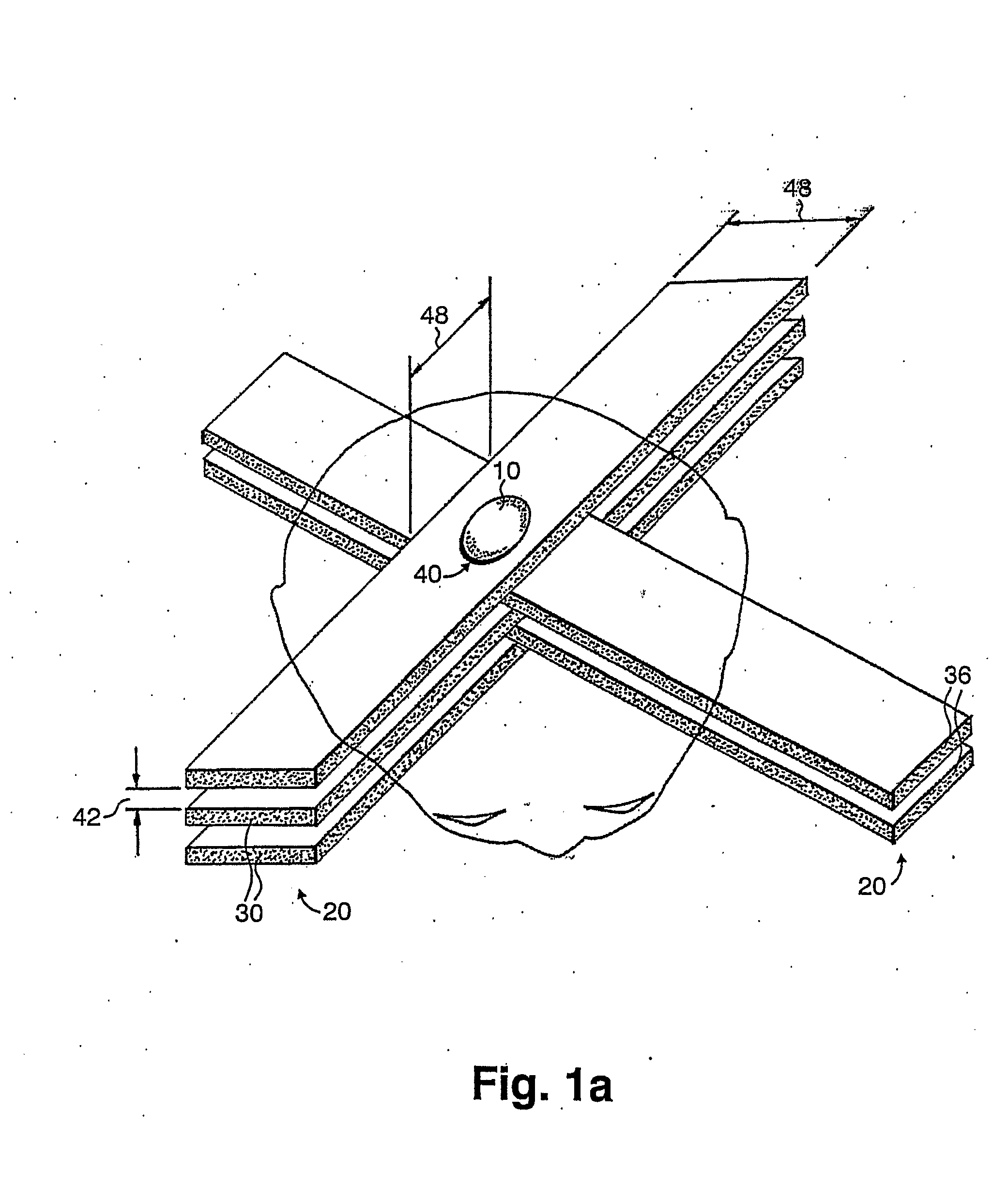
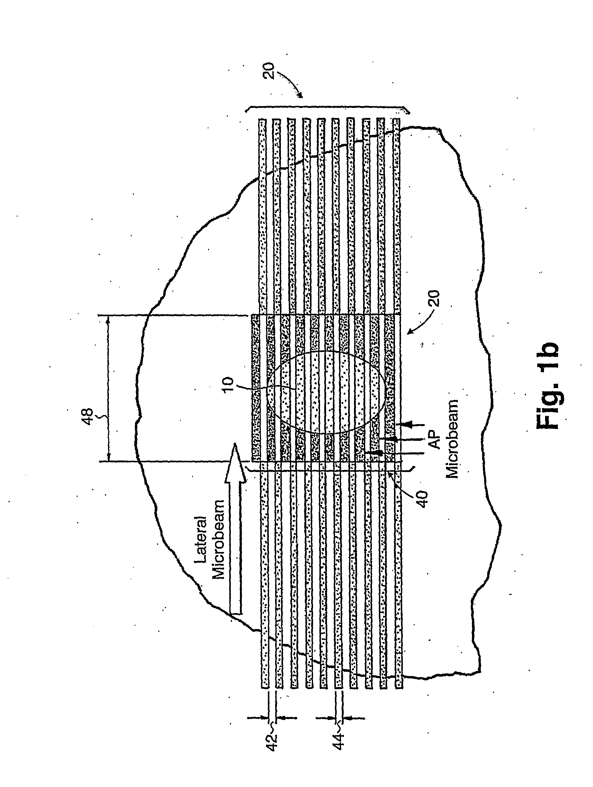
[0018]The present invention, which addresses the needs of the prior art, relates to more efficient methods of radiation therapy which greatly enhance the therapeutic dose and damage to target tissue, such as a tumor, while simultaneously reducing damage to normal tissue in the path of the irradiating beam. This result is achieved by providing a different type of radiation, i.e., a broad beam effect, to the tumor than to the normal tissue in the beam path.
[0028]Any of the interlaced MRT techniques of the present invention, e.g., BIMRT and MIMRT, may further include providing a concentration of a radiation contrast agent substantially only to the target tissue for preferential X-ray absorption. The concentration enhances an in-beam dose of the high energy electromagnetic radiation in each of the at least two parallel, spatially distinct microbeams of the at least two non-intersecting arrays interleaved substantially only within the target tissue.
[0032]In the second technique of the present invention, a therapeutic dose of high energy electromagnetic radiation is delivered substantially only to a target tissue by generating a broad beam radiation effect only within the target tissue. The act of delivering includes irradiating the target tissue with at least one array of microbeams having at least two parallel, spatially distinct microbeams. The method further includes providing a concentration of a radiation scattering agent substantially only to the target tissue. The radiation scattering agent scatters the high energy electromagnetic radiation in a substantially perpendicular direction to an irradiation direction of the individual microbeams, thus raising a valley dose, i.e., the dose between each of the at least two parallel, spatially distinct microbeams, substantially only within the target tissue. The raising of the valley dose between microbeams in the array relative to the in-beam dose provides the broad beam effect substantially only within the target tissue.
[0040]As a result, the present invention provides more efficient methods of radiation therapy by employing microbeams in particular geometries, including BIMRT and MIMRT, or by using microbeam array(s) in combination with a radiation scattering agent to produce a broad beam effect only within a target tissue. The methods may include the use of contrast agents, which are preferentially up-taken by the tumor tissue, of two different types: (a) those including heavy elements to enhance in-beam absorption of microbeam radiation, preferably used with the interlaced technique of the present invention, e.g., BIMRT and MIMRT; and (b) those including lighter elements to produce scattering of microbeam radiation, preferably used with non-interleaving microbeams to preferentially increase the valley dose within the target tissue. Both types of agents will greatly enhance the therapeutic dose and contribute to a broad beam effect at the site of the tumor. Safe doses are maintained to normal tissue in the path of the irradiating beam by the particular geometries of irradiation provided using microbeams.
[0041]The present invention, which addresses the needs of the prior art, also relates to more efficient methods of radiation therapy which greatly enhance the therapeutic dose and damage to selected tissue types in a target volume of the central nervous system or other organ affected by a disease or condition. Simultaneously, damage to surrounding normal tissue in the path of the irradiating beam is minimized. This result is achieved by providing a broad beam effect substantially only within the target volume using microbeam arrays.
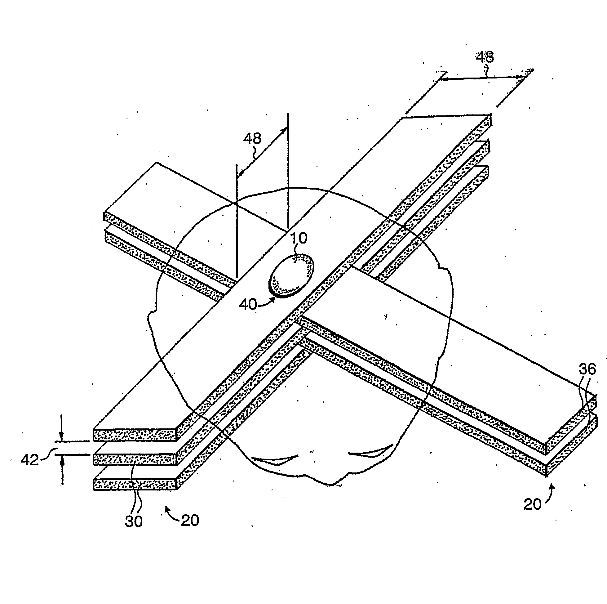
[0042]The present invention further relates to a method of performing radiation therapy on a subject suffering from a disease or condition. The method includes delivering a dose of high energy electromagnetic radiation to selected tissue in a target volume in an amount sufficient to damage or ablate at least a portion of the selected tissue without inducing permanent damage to tissue external to the target volume by generating a broad beam radiation effect only within the target volume. The delivering step includes irradiating the selected tissue with at least two arrays of microbeams, where each array includes at least two parallel, spatially distinct microbeams, and interleaving the at least two arrays at the target volume to form a substantially continuous broad beam of radiation within the selected tissue in the target volume defined by the interleaved microbeams.

Problems solved by technology

Conventional treatments such as surgery, chemotherapy and radiation therapy have exhibited favorable results in many cases, while failing to be completely satisfactory and effective in all instances.
For example, the effectiveness of orthodox radiation therapy on deep pulmonary, bronchial, and esophageal tumors is limited by the risk of radiation pneumonitis.
This goal is particularly difficult to achieve in treating central nervous system (CNS) tumors.
The survival statistics of patients with high grade gliomas in the brain, or lower grade gliomas and metastatic tumors in the spinal cord have not improved appreciably in recent years using conventional surgical techniques and conventional radiotherapy.
The doses that can be delivered to malignant CNS tumors are limited by the tolerance of normal brain and spinal cord to radiation.
For lower grade CNS tumors, the ratio of radiotherapy doses that produce normal CNS toxicity and those that control the tumor is so close that it often renders radiotherapy ineffective, or results in neurological complications from radiotoxicity to the normal CNS surrounding the tumor.
These sensitizers, however, are commonly highly toxic, and care must be taken not to administer too large of a quantity of these compounds to a subject.
Even with careful administration, an unwanted risk to the subject is imposed by this method, because of variations in tolerance levels among subjects.
In practice, however, there is still the risk of radiation-induced tissue necrosis by the broad X-ray beam.
It is extremely toxic, however, and has a very small amount of gadolinium in it.

Method used


Image

  • Methods for Implementing Microbeam Radiation Therapy
  • Methods for Implementing Microbeam Radiation Therapy
  • Methods for Implementing Microbeam Radiation Therapy

Examples


example 1

[0223]The following study was carried out at the National Synchrotron Light Source (NSLS), Brookhaven National Laboratory, Upton, N.Y., 11973. The results show the efficacy of gold nanoparticles combined with BIMRT. Mice with subcutaneous murine mammary carcinoma tumor EMT-6 tumor inoculated behind their neck were treated with the BIMRT of the present invention. The microbeam arrays had a 0.68 millimeters (mm) beam thickness and 1.36 mm center-to-center beam spacing, i.e., 0.68 mm inter-beam spacing. The gold nanoparticles used in the study were about 1.9 nm in diameter. At the ninth day of inoculation, when the tumor sizes averaged about 100 mm3, the mice were randomized in five groups of seven (7) mice each for the following treatments: Group A: 55 Gy BIMRT; Group B: 55 Gy broad beams (bidirectional, 2×27.5 Gy); Group C: 35 Gy BIMRT; Group D: 35 Gy BIMRT with gold nanoparticles; and Group E: Unirradiated controls. The gold nanoparticles, 0.2 ml in volume, were injected via the tai...

example 2

[0226]The major challenges in the use of x-ray tubes for microbeam radiation therapy are the divergence of the beams of most x-ray sources (with the exception of synchrotron sources) in the direction perpendicular to the microplanar beams, and the relatively larger source spot size. To optimize the dose radiation profile of the microbeams using conventional x-ray tubes, it is preferable to use a thick beam of about 0.7 mm. In addition, irradiation using a single microplanar beam at a time is also preferable, as demonstrated in the following example.

[0227]In this example, the radiation source is an x-ray tube with a source spot size of 0.4 mm in the direction perpendicular to the planes of the microbeams. The source is positioned 1 meter away from the slit that forms the microplanar beam. The slit is positioned 25 cm from the center of the target. The beam's thickness is 0.7 mm, leading to a nominal beam spacing on-center of 1.4 mm (i.e., inter-beam spacing of 700 microns). The targe...


PUM


Abstract

A method of performing microbeam radiation therapy (MRT) includes delivering a dose only to selected tissue in a target volume (10) with continuous broad beam, first, by interleaving arrays of microplanar beams (30,36) only at the target (10). Administered contrast agents can supplement the effect by preferentially increasing the target dose relative to dose in normal tissue. A broad beam effect is alternatively created using non-interleaving microbeam array(s) with scattering agents administered to selected tissue that preferentially increase valley dose (69) within target to approximate broad beam. The methods of interleaving microbeams are also applied to treat diseases and conditions by ablating at least a portion of selected tissue, or by damaging blood-brain barrier for efficient drug and / or cell administration. A system for performing interlaced microbeam radiosurgery preferably includes two orthogonal radiation source arms (102) for producing and interleaving microbeam arrays (30,36) at the target volume (10). The methods treat tumors, pain, epilepsy, and neurological diseases.

Description

CROSS REFERENCE TO RELATED APPLICATIONS[0001]This application is a continuation-in-part application of pending U.S. patent application Ser. No. 11 / 054,001, filed Feb. 10, 2005, which is incorporated herein by reference in its entirety.STATEMENT OF GOVERNMENT LICENSE RIGHTS[0002]This invention was made with Government support under contract number DE-AC02-98CH10886, awarded by the U.S. Department of Energy. The Government has certain rights in the invention.FIELD OF THE INVENTION[0003]The present invention relates generally to methods for performing microbeam radiation therapy on a subject for treatment of tumors and of diseases and conditions affecting the central nervous system and other organs, and more particularly to methods of using microbeam arrays to produce a broad beam effect only within a target volume, for example, within a tumor, thus increasing the therapeutic effect of microbeam radiation therapy.BACKGROUND OF THE INVENTION[0004]Cancer continues to be one of the foremo...

Claims


Application Information

Patent Timeline
IPC IPC(8): A61N5/10
CPCA61N2005/1091A61N5/1045A61N2005/1098A61N2005/109
Inventor DILMANIAN, F. AVRAHAMMORRIS, GERARD M.HAINFELD, JAMES
Owner BROOKHAVEN SCI ASSOCS
Learn More